Tag Archives: mesentery

Bucket Handle Injury – Part 2

Yesterday, I wrote about the basics of bucket handle injuries of the intestine. Today, I’ll deal with diagnosing them.

An understanding of the mechanism of injury and a good physical exam are paramount. If the patient took a significant blow to the abdomen, especially in a motor vehicle crash (lap belt), be very suspicious. Any abdominal pain is of concern, particularly in the right lower quadrant (most common injury location). If a CT is indicated and there are focal changes in the mesenery or bowel wall, a trip to the OR is advised.

In some patients, the bowel is devascularized and takes 2-3 days to become necrotic. They experience slowly increasing focal pain, and once this develops it’s time to go to the operating room.

Intubated and/or comatose patients can be problematic in making this diagnosis. There is no physical exam, so the trauma professional has to rely on surrogates. The white blood cell (WBC) count is very helpful. The WBC count is typically elevated into the 15,000-20,000 range immediately after trauma, and declines to normal within about 12 hours. If it begins to climb again after 24 hours, especially if it exceeds 20,000, an intestinal injury is likely.

CT scan and abdominal ultrasound are also helpful. A repeat CT scan may show a change in the volume of fluid, or a change in its character. If the amount of fluid increases significantly, or if a fluid bi-layer is seen, a bucket handle injury is very likely. These findings are pertinent in awake patients as well, but the physical exam usually makes use of these diagnostics unnecessary.

Related posts:

Bucket Handle Injury – Part 1

Bucket Handle Injury

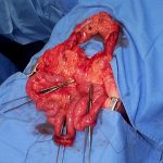
A bucket handle injury is a type of mesenteric injury of the intestine. The intestine itself separates from the mesentery, leaving a devascularized segment of bowel that looks like the handle on a bucket (get it?).

These injuries can occur after blunt trauma to the abdomen. The force required is rather extreme, so the usual mechanism is motor vehicle crash. In theory, it could occur after a fall from a significant height, and I have seen once case where a wood fragment was hurled at the abdomen by a malfunctioning lathe.

The mechanics of this injury are related to fixed vs mobile structures in the abdomen. Injuries tend to occur adjacent to areas of the intestine that are fixed, such as the cecum, ligament of Treitz, colonic flexures and rectum. During sudden deceleration, portions of the intestine adjacent to these areas continue to move, pulling on the nearby attachments. This causes the intestine itself to pull off of its mesentery.

The terminal ileum is the most common site for bucket handle tears. Proximal jejunum, transverse colon, and sigmoid colon are other possible areas. The picture above shows multiple bucket handle injuries in one patient. There are 3 injuries in the small bowel, and one involving the entire transverse colon. Note the obviously devascularized segment at the bottom center of the photo.

Always think about the possibility of this injury in patients with very high speed decelerations. Seat belt marks have a particularly high association with this injury. If your patient has an abnormal exam in the right lower quadrant, or if the CT shows unusual changes there (“dirty” mesenteric fat, thickened bowel wall, extravasation), I recommend a trip to the OR. In these cases, an injury will nearly always be present.

Tomorrow: These injuries can be subtle in an awake patient with a reliable exam. On Friday I’ll write about how you can detect it in unconscious patients.

Source: personal archive. Not treated at Regions Hospital

Identifying Bowel and Mesenteric Injury by CT

CT scan is an invaluable tool for evaluating blunt abdominal trauma. Although it is very good at detecting solid organ injury, it is not so great with intestinal and mesenteric injuries. Older studies have suggested that CT can detect mesenteric injuries if done right, but a newly published study has shown good accuracy with a few imaging tweaks.

A Taiwanese study looked at a series of prospectively studied victims of blunt abdominal trauma. Patients with abdominal pain or a positive FAST were entrolled (total 106). IV contrast was given, and scans during the arterial, portal, and equilibrium contrast phases were performed using a multidetector scanner. Images were read in a blinded fashion.

A total of 13 of 23 patients who underwent laparotomy were found to have a bowel or mesenteric injury. Five had bowel injury, 4 had mesenteric hemorrhage, and 4 had both. Mesenteric contrast extravasation was seen in 7 patients, and this correlated with mesenteric bleeding at laparotomy.

The authors found that the following signs on CT scan indicated injury:

  • Full or partial thickness change in bowel wall appearance
  • Increased mesenteric density
  • Free fluid without solid organ injury

Bottom line: This study shows that CT scan can detect bowel and mesenteric injury reliably if you scan the patient 3 times! This seems like over-radiation and overkill. A more intelligent way to approach this would be to perform a normal trauma abdominal scan. If a suspicious area of mesenteric or bowel thickening is seen, then a limited rescan through the affected area only for equilibrium phase images may be warranted. If actual contrast extrvasation is seen, no further scanning is needed. A quick trip to the OR is in order.

Reference: Contrast-enhanced multiphasic computed tomography for identifying life-threatening mesenteric hemorrhage and transmural bowel injuries. J Trauma 71(3):543-548, 2011.