Tag Archives: liver

Contrast Blush in Children

A contrast blush is occasionally seen on abdominal CT in patients with solid organ injury. This represents active arterial extravasation from the injured organ. In most institutions, this is grounds for call interventional radiology to evaluate and possibly embolize the problem. The image below shows a typical blush from extravasation.

Splenic contrast blush

This thinking is fairly routine and supported by the literature in adults. However, it cannot be generalized to children!

Children have more elastic tissue in their spleen and tend to do better with nonoperative management than adults. The same holds true for contrast blushes. The vast majority of children will stop bleeding on their own, despite the appearance of a large blush. In fact, if children are taken to angiography, it is commonplace for no extravasation to be seen!

Angiography introduces the risk of local complications in the femoral artery as well as more proximal ones. That, coupled with the fact that embolization is rarely needed, should keep any prudent trauma surgeon from ordering the test. It should be reserved for cases where nonoperative management is failing, but hypotension (hard fail) has not yet occurred.

The only difficult questions is “when is a child no longer a child?” Is there an age cutoff at which the spleen starts acting like an adult and keeps on bleeding? Unfortunately, we don’t know. I recommend that you use the “eyeball test”, and reserve angiography for kids with contrast extravasation who look like adults (size and body habitus).

Reference: What is the significance of contrast “blush” in pediatric blunt splenic trauma? Davies et al. J Pediatric Surg 2010 May; 45(5):916-20.

Pop Quiz: Interesting Case!

A 16 year old male was thrown against the handlebars during a motorcycle crash at about 40 mph. He dusted himself off and went home for a few hours. Unfortunately, he slowly developed some abdominal pain.

He presented to an ED several hours later. He was found to have mild, diffuse abdominal pain, normal vital signs, and a positive abdominal FAST exam. CT scan showed a grade IV spleen injury and a grade II liver injury in the right lobe with no extravasation or pseudoaneurysm noted. He was successfully treated nonoperatively and was sent home.

One month later he returns to the ED complaining of a single episode of hematochezia (approximately 200cc). He has an entirely normal exam and vital signs.

Here are my questions for you:

  • Was the initial management appropriate?
  • Should anything additional have been done during the first admission?
  • What is the diagnosis now?
  • What diagnostic or therapeutic maneuvers are indicated now?

Please tweet your guesses, or leave comments below. Hints tomorrow and answers on Friday. Good luck!

Patient not treated at Regions Hospital

Trauma Residents: How To Remember Liver Anatomy

In trauma surgery, operative management of liver injury is usually messy business, with little time for nice anatomic resections. However, an understanding of the basic anatomy, especially that of the vascular supply is crucial for saving your patient.

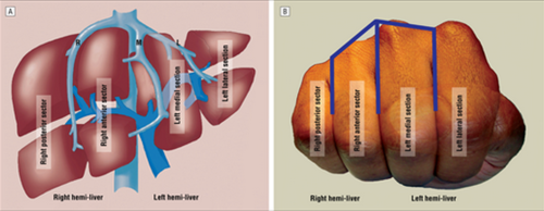
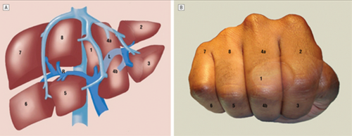
A cool tool for remembering Couinaud’s segments and the overall layout of liver anatomy was published in the Archives of Surgery recently. It makes use of a model, which consists of your hand! Just make a fist with your right hand and tuck the thumb behind the other fingers.


The fingers can then be numbered according to the Couinaud segments, with the caudate lobe (segment 1) represented by the thumb that is tucked away. The PIP joints represent the plane that the portal vein runs through, with branches going to upper and lower segments. Note how the ring finger normally lies a little more anterior than the little finger in this position, just like the sectors of the right lobe.

The creases between the fingers represent the left, middle and right hepatic veins.


The right hepatic vein is located between the right anterior and posterior sectors and the left hepatic vein sits between the left medial and lateral sectors. The middle hepatic vein is in between the left and right hemi-liver.

Bottom line: This “handy” liver model is available immediately in the OR and is already sterile. It can help visualize liver structures that may be injured quickly and accurately to speed your operative approach to the problem.

Reference: A Handy Tool to Teach Segmental Liver Anatomy to Surgical Trainees. Arch Surg 147(8):692-693, 2012.

DVT Prophylaxis After Solid Organ Injury

Nonoperative management of solid organ injury is the norm, and has reduced the operative rate significantly. At the same time, the recognition that development of deep venous thrombosis (DVT) in trauma patients is commonplace creates uncertainty? Is it safe to give chemical prophylaxis with low molecular weight heparin (LMWH)? How soon after injury?

The trauma group at USC+LAC published the findings of a retrospective review of 312 patients undergoing nonoperative management for their liver, spleen or kidney injuries. They looked at chemical prophylaxis administration and its relationship to failure of nonop management of solid organ injury.

As expected, as the grade of the solid organ injury increased, so did the failure rate of nonoperative management. Administration of low molecular weight heparin, such as enoxaparin, did not increase failure rate in this study. All but one failure occurred in patients who had not yet received the injections. Likewise, two DVT and two pulmonary embolisms occurred, but only in patients who had not yet received prophylaxis. 

Bottom line: This small study offers some assurance that early prophylaxis is okay, and a few prospective studies do exist. UCSF / San Francisco General is comfortable beginning chemical prophylaxis 36 hours postop, regardless of solid organ injury. Look for more guidance on this issue in the near future. Until then, consider starting LMWH prophylaxis early to avoid complications from DVT or PE.

Reference: Thromboembolic prophylaxis with low-molecular-weight heparin in patients with blunt solid abdominal organ injuries undergoing nonoperative management: current practice and outcomes. J Trauma 70(1): 141-147, 2011.

Algorithm For Nonoperative Management of Blunt Hepatic Trauma

Yesterday, I posted the Western Trauma Association’s algorithm for operative management of blunt liver trauma. Click here to view it. Today, I’m going to discuss their algorithm for nonoperative management. 

The algorithm is fairly self-explanatory. Click on the image above to read the annotated text for details on each step. Note: this requires full access to the Journal of Trauma.

Some key points in this algorithm:

  • Unstable patients need rapid identification of the cause. If the FAST is positive ©, then you need to go to the OR and use the operative algorithm.
  • CT scan is used for diagnosis in stable patients (F), but if a liver injury is seen and they become unstable at any time, go to the OR.
  • Contrast extravasation in a stable patient should prompt an evaluation and possible embolization by interventional radiography (G).
  • If complications develop (SIRS, abdominal pain, fever, jaundice), a repeat CT is indicated (K).
  • Abscesses and focal collections of bile may be managed by interventional radiology (L,M). Persistent bile leak may be decreased by ERCP and sphincterotomy (O).
  • Bile ascites or large hemoperitoneum may be managed using laparoscopy with drainage (N).

Reference: Western Trauma Association critical decisions in trauma: nonoperative management of adult blunt hepatic trauma. J Trauma. 67:1144–1148, 2009.