Using A 3D Printer To Plan Orthopaedic Surgery

I’ve previously written about new printing technology applications in trauma. A recent article details a new way to use 3D printing technology for planning complex orthopaedic procedures.

An orthopedic registrar in Monklands Hospital (North Lanarkshire, Scotland) found a way to combine new printing technology and orthopaedics. CT scans are routinely taken of complex fractures. Scanners now have powerful software that enables us to create 3D reconstructions from the helical or axial images. However, these are just a series of 2D images viewed on a computer monitor.

Mr. Mark Frame found a way to convert the CT information into a format that can be used as input for a 3D printer. Using two open source (free) software packages for the Mac, OsiriX and MeshLab, he was able to create a medical quality 3D image file. The file was sent to a company that printed it using a 3D printer.

The cost? About $235 US plus a little time for a complete model of the pelvis. The advantage? The actual size 3D model can be used to select hardware and practice the repair technique. And the cost to own a 3D printer keeps coming down!

Related posts:

What’s The INR of Fresh Frozen Plasma?

So what’s the INR of FFP? Or stated another way, what’s the lowest you can correct a patient’s INR using infusions of fresh frozen plasma?

One of the mainstays of correcting coagulopathy, either from hemorrhage or due to medication like warfarin, is transfusion of FFP. Frequently, clinicians will write orders to administer FFP until a certain INR is achieved. What is a reaonable INR?

A “normal” INR is 1.0, plus or minus about 0.2, depending on your laboratory. However, two separate studies have shown that transfusion of FFP will not reliably decrease the INR below about 1.7. 

Bottom line: The answer to the question is about 1.6. If any clinician orders FFP transfusions with a goal INR below this, it probably won’t happen. And since transfusions of any product have risks, my “juice to squeeze” ratio of risk vs benefit begins to fail at an INR of 1.6. Below that point, the patient needs a normal temperature and good perfusion to drop their INR further.

References:

  • Toward rational fresh frozen plasma transfusion: the effect of plasma transfusion on coagulation test results. Am J Clin Pathol 126(1):133-139, 2006.
  • Effect of fresh frozen plasma transfusion on prothrombin time and bleeding in patients with mild coagulation abnormalities. Transfusion 46(8):1279-1285, 2006.

Lab Values From Intraosseous Blood

The intraosseous access device (IO) has been a lifesaver by providing vascular access in patients who are difficult IV sticks. In some cases, it is even difficult to draw blood in these patients by a direct venipuncture. So is it okay to send IO blood to the lab for analysis during a trauma resuscitation?

A study using 10 volunteers was published last year (imagine volunteering to have an IO needle placed)! All IO devices were inserted in the proximal humerus. Here is a summary of the results comparing IO and IV blood:

  • Hemoglobin / hematocrit – good correlation
  • White blood cell count – no correlation
  • Platelet count – no correlation
  • Sodium – no correlation but within 5% of IV value
  • Potassium – no correlation
  • Choloride – good correlation
  • Serum CO2 – no correlation
  • Calcium – no correlation but within 10% of IV value
  • Glucose – good correlation
  • BUN / Creatinine – good correlation

Bottom line: Intraosseous blood can be used if blood from arterial or venous puncture is not available. Discarding the first 2cc of marrow aspirated improves the accuracy of the lab results obtained. The important tests (hemoglobin/hematocrit, glucose) are reasonably accurate, as are Na, Cl, BUN, and creatinine. The use of IO blood for type and cross is not yet widely accepted by blood banks, but can be used until other blood is available.

Related post:

Reference: A new study of intraosseous blood for laboratory analysis. Arch Path Lab Med 134(9):1253-1260, 2010.

Identifying Bowel and Mesenteric Injury by CT

CT scan is an invaluable tool for evaluating blunt abdominal trauma. Although it is very good at detecting solid organ injury, it is not so great with intestinal and mesenteric injuries. Older studies have suggested that CT can detect mesenteric injuries if done right, but a newly published study has shown good accuracy with a few imaging tweaks.

A Taiwanese study looked at a series of prospectively studied victims of blunt abdominal trauma. Patients with abdominal pain or a positive FAST were entrolled (total 106). IV contrast was given, and scans during the arterial, portal, and equilibrium contrast phases were performed using a multidetector scanner. Images were read in a blinded fashion.

A total of 13 of 23 patients who underwent laparotomy were found to have a bowel or mesenteric injury. Five had bowel injury, 4 had mesenteric hemorrhage, and 4 had both. Mesenteric contrast extravasation was seen in 7 patients, and this correlated with mesenteric bleeding at laparotomy.

The authors found that the following signs on CT scan indicated injury:

  • Full or partial thickness change in bowel wall appearance
  • Increased mesenteric density
  • Free fluid without solid organ injury

Bottom line: This study shows that CT scan can detect bowel and mesenteric injury reliably if you scan the patient 3 times! This seems like over-radiation and overkill. A more intelligent way to approach this would be to perform a normal trauma abdominal scan. If a suspicious area of mesenteric or bowel thickening is seen, then a limited rescan through the affected area only for equilibrium phase images may be warranted. If actual contrast extrvasation is seen, no further scanning is needed. A quick trip to the OR is in order.

Reference: Contrast-enhanced multiphasic computed tomography for identifying life-threatening mesenteric hemorrhage and transmural bowel injuries. J Trauma 71(3):543-548, 2011.

There’s An App For That: Reading Xrays on Your iPhone/iPad

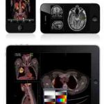
MIM Software has developed an app that allows trauma professionals to view xrays on their mobile device. The app actually received FDA clearance for use earlier this year, and is available as a free download.

The catch is that the software only works with MIM’s cloud-based product for managing radiographic images, called MIMcloud. It is primarily intended for those who do not have access to a full image viewing workstation (MIMfusion).

Home of the Trauma Professional's Blog

Do you want to get a daily email every time there’s a new post? See what I’m up to.

Click here to get details and subscribe!

[accua-form fid=”1″]

[mc4wp_form id=”2023″]