Helicopter EMS: The Risks

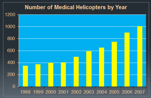
Yesterday, I wrote about the (unclear) benefits of helicopter EMS transports. Today, I’ll cover the risks. The number of medical helicopters in the US has grown dramatically since 2002.

image

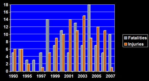
As can be expected, the number of mishaps should go up as well. 

image

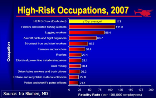
Although it looks like the fatal and injury accidents peaked and then declined, it does not look as good when compared to the rest of the aviation industry. Consequently, being on a helicopter EMS (HEMS) crew has become one of the more dangerous professions.

image

And unfortunately, the numbers have not improved much during the past five years. So what to do? Make it a big PI project. Approach it systematically, analyze the issues, and create some guidelines and protocols for all to follow.

Tomorrow, I’ll review new guidelines for HEMS released by the American College of Surgeons Committee on Trauma.

Related posts:

Reference: Medical helicopter accidents in the United States: a 10 year review. J Trauma 56:1325-1329, 2004.

Helicopter EMS: The Benefits?

The use of medical helicopters has grown at an astonishing rate in the 10+ years since Medicare got involved with payment for this service. All high level trauma centers have helicopter landing facilities, and many either own or are a part owner in at least one helicopter EMS service (HEMS).

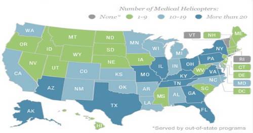
Here’s a state by state breakdown of the number of medical helicopters:

image

It’s gotten to the point where the indication for summoning a HEMS service seems to be the presence of a patient to ride on it! 

A lot of papers have been published in the past 20 years trying to justify the benefits of using these services. As is the usually case when a lot of papers are published on one subject, most of them are not very good. Lots of studies have been performed to try to justify their use, and most were not successful. The following items have been scrutinized:

  • Interfacility transfers
  • Trauma
  • Pediatric transfers 
  • Pediatric trauma
  • Burns
  • OB
  • Neonatal
  • Rural trauma

Most of these papers found little, if any, benefit. The ones that did tended to be published by institutions that owned these services, raising the question of bias. The one thing that was always significantly different was the cost. HEMS costs at least 10 times more that ground EMS transport.

So the benefits are not very clear. What about the risks? I’ll talk about those tomorrow.

Click here to view the interactive state map of medical helicopters. See where your state is with respect to number of ships and services, and how busy they are.

Related posts:

The Medical Orthopaedic Trauma Service

Our population is aging, and falls continue to be a leading cause of injury and morbidity in the elderly. Unfortunately, many elders have significant medical conditions that make them more likely to suffer unfortunate complications from their injuries and the procedures that repair them.

A few hospitals around the world are applying a more multidisciplinary approach than the traditional model. One example is the Medical Orthopaedic Trauma Service (MOTS) at New York-Presbyterian Hospital/Weill Cornell Medical Center. Any elderly patient who has suffered a fracture is seen in the ED by both an emergency physician and a hospitalist from the MOTS team. Once in the hospital, the hospitalist and orthopaedic surgeon try to determine the reason for the fall, assess for risk factors such as osteoporosis, provide comprehensive medical management, provide pain control, and of course, fix the fracture. 

This medical center recently published a paper looking at their success with this model. They retrospectively reviewed 306 patients with femur fractures involving the greater trochanter. They looked at complications, length of stay, readmission rate and post-discharge mortality. No change in length of stay was noted, but there were significantly fewer complications, specifically catheter associated urinary tract infections and arrhythmias. The readmission rate was somewhat shorter in the MOTS group, but did not quite achieve significance with regression analysis.

Bottom line: This type of multidisciplinary approach to these fragile patients makes sense. Hospitalists, especially those with geriatric experience, can have a significant impact on the safety and outcomes of these patients. But even beyond this, all trauma professionals need to look for and correct the reasons for the fall, not just fix the bones and send our elders home. This responsibility starts in the field with prehospital providers, and continues with hospital through the entire inpatient stay.

Related post:

Reference: The medical orthopaedic service (MOTS): an innovative multidisciplinary team model that decreases in-hospital complications in patients with hip fractures. J Orthopaedic Trauma 26(6):379-383, 2012.
Trauma 20 Years Ago: CAVR For Hypothermia

Hypothermia is the bane of major trauma resuscitation, causing mortality to skyrocket. A number of rewarming techniques have been developed over the years. These are classified as passive (the patient generates their own heat) or active (we deliver calories to them), and noninvasive vs invasive. Rewarming speed increases as we move from passive to active and from noninvasive to invasive.

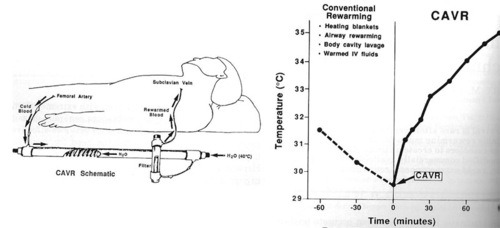
Continuous arteriovenous rewarming (CAVR) is one of the invasive techniques used today. Its use in humans was first reported 20 years ago this month. Larry Gentilello at Harborview in Seattle had experimented with this technique in animals, and reported one case of use in a human who had crashed his car into icy water. After a 20 minute extrication, the patient was pulseless with fixed and dilated pupils, but he regained pulse and blood pressure at the hospital.

The initial core temperature was 31.5C. Peritoneal, bladder and gastric lavage were carried out for warming, as was delivery of warm inspired gas via the ventilator. However, after an hour the temperature had dropped to 29.5C. CAVR was initiated as a last-ditch effort using a jerry-rigged Rapid Fluid Warmer from Level 1 Technologies. The core temperature was raised to 35C after 85 minutes.

The patient did have typical complications (ARDS, acute renal failure), but survived with recovery of his renal and pulmonary function, and a normal neurologic exam. At the time, the authors were unsure whether the complications were due to the near-drowning or the rapid rewarming.

Reference: Continuous arteriovenous rewarming: report of a new technique for treating hypothermia. J Trauma 31(8):1151-1154, 1991.

Related posts:

Rapid Noninvasive Rewarming Using a Hubbard Tank

It’s that time of year again in Minnesota. We’re getting 5 inches of snow tonight, so hypothermia season is officially here! I’m republishing a technique for rewarming patients faster than just about any other method. Most burn centers have large tanks for handling burn wounds, and many hospital have smaller therapy tanks that can be used for the same purpose.

Hypothermic patients need to be rewarmed using the most appropriate method. Patients with mild hypothermia (32-35 degrees centigrade) generally only require removal of wet clothing and surface warming. Moderate hypothermia (28-32 degrees C) to severe hypothermia (<28 degrees C) is very serious and requires more aggressive central rewarming techniques.

Basic central rewarming techniques, such as warm inspired gases, warm IV fluids, and gastric or peritoneal lavage can raise the temperature about 3 degrees per hour. 

Rapid central rewarming techniques, like thoracic lavage (6 degrees/hr), AV bypass devices (1-4 degrees/hr), and cardiopulmonary bypass (18 degrees/hr) are typically used on patients with severe hypothermia.

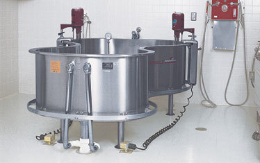
A technique that we use at Regions Hospital involves the use of the Burn Center’s Hubbard Tank. Patients are carefully immersed, torso first, then one extremity at a time to avoid rebound hypothermia. It is possible to increase core temperature using this method faster than bypass (>20 degrees centigrade/hr)! Typical time in the tank is an hour or less for any degree of hypothermia.

Patients can be immersed with EKG monitors and IV lines in place. Temperature monitoring should be performed using a thermistor tipped urinary catheter. Many hospitals don’t have a full Hubbard tank, but do have smaller therapy baths that work nearly as well.

Tips:

  1. A physician must stay with the patient while immersed in case arrhythmias develop.
  2. Position the urinary catheter and collecting bag in such a way that urine in the tubing does not backwash into the bladder. This will falsely and rapidly increase the temperature reading.

Related post:

Home of the Trauma Professional's Blog

Do you want to get a daily email every time there’s a new post? See what I’m up to.

Click here to get details and subscribe!

[accua-form fid=”1″]

[mc4wp_form id=”2023″]