All posts by The Trauma Pro

What Is: A Rubber Bullet?

The protests in cities across the country continue. Many are peaceful, but not all. In some cases, police have resorted to “non-lethal” weapons to control and disperse crowds.

Although these weapons are called non-lethal, that’s not entirely true. The projectiles, gases, and powders that are being used all have some degree of morbidity and mortality. They are certainly less so than traditional projectiles (bullets), but serious and fatal injures can and do occur.

One item that is talked about in the news is the rubber bullet. What are these, exactly? The generic term is a “kinetic impact projectile” (KIP). It encompasses a variety of objects that are not designed to penetrate flesh like a regular bullet. They can be bullets, beanbags, sponges, pellets, and other odds and ends.

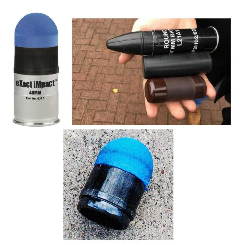
And the so-called “rubber bullet” isn’t even necessarily made of rubber. It can be plastic, metal, rubber, or other substances.

There is very little published data on injuries caused by KIPs. Because of their odd shapes, they tend to tumble when they are fired. This decreases aiming accuracy substantially when the target is distant. They are designed to be aimed at the lower extremities. However, if the aim is too high or the round is fired at close range, it can be lethal.

Here are some typical injuries that have been descirbed:

  • Subdural and intraparenchymal hematomas
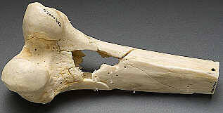
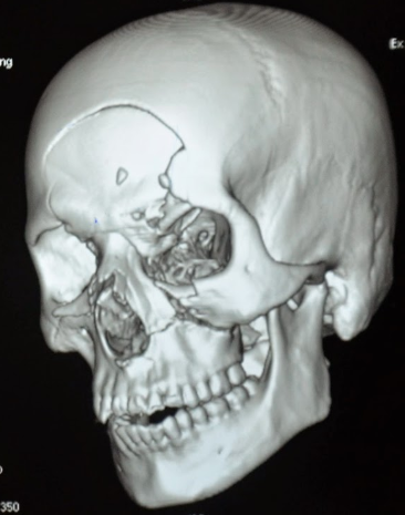
  • Skull and facial fractures
  • Eye injuries leading to blindness (this happened to a photographer in Minneapolis last week)
  • Rib fractures and pulmonary contusions
  • Spleen laceration
  • Blunt intestinal injury

Here’s a video from one of the manufacturers that shows the amount of target deformation caused by a sponge tipped bullet. Very impressive.

YouTube player

(Tumblr viewers please click here to view video)

Bottom line: Although they sound relatively innocuous, kinetic impact projectiles of any kind are far from it. If you are called to treat a patient who has been shot with one, be sure to do a very thorough evaluation. Most head, neck, and torso injuries should undergo CT scanning to delineate deep or occult injuries in detail. In-hospital observation of torso injuries is warranted, as well as a good tertiary exam.

More On Lead Poisoning And Retained Bullets

Trauma professionals frequently have to leave bullets in patients. It is often more disruptive to go digging the projectiles out than to just leave them in place. But patients always want to know why and what the consequences might be.

In my last post, I discussed a very old paper on what we know about lead levels and retained bullets. Very recently, a meta-analysis was published that provides a better picture of this topic. They somehow managed to find over 2000 articles dealing with lead toxicity and bullets out there. But after someone had the pleasure of reviewing each of them, they found only 12 that had any meaningful or actionable information.

Here are the factoids:

  • All studies were observational (duh! It would be difficult to get your IRB to approve a study where patients were shot on purpose)
  • There were five cross-sectional studies, four case-control studies, and three prospective cohort studies
  • The studies were small, with a median of only 26 patients (range 15-120)
  • Eleven of the twelve studies showed an association with retained bullets and elevated blood lead levels
  • Three studies showed elevated blood levels if a fracture was present
  • The higher the number of retained fragments, the more likely lead levels were to be high
  • Higher lead levels were associated with retained fragments near a bone or joint
  • There were no good correlations with number of fragments and location vs actual lead toxicity

Bottom line: Even using meta-analysis, it is difficult to tease out meaningful answers to this question. That speaks to the low numbers of papers and their quality. However, this study does provide a little bit of guidance.

Retained bullet fragments are probably not a big worry in most patients. The bothersome cases are those where the fragments are in or near a bone or joint. And even though few patients actually developed lead toxicity, lead levels approaching 5 micrograms/dL can have physiologically significant negative effects. 

Recommendation: If your patient has a retained bullet fragment near a bone or joint, or they have “multiple” retained fragments (no good definition of this), they should have blood lead levels measured every three months for a year. If the level is rising, and certainly if it reaches the 5μ/dL level, attempts should be made to remove the fragments.

Reference: Lead toxicity from retained bullet fragments: A systematic review and meta-analysis. J Trauma 87(3):707-716, 2019.

Can Lead Poisoning Occur After A Gunshot?

This is a fairly common question from victims of gunshots and their families. As you know, bullets are routinely left in place unless they are superficial. It may cause more damage to try to extract one, especially if it has come to rest in a deep location. But is there danger in leaving the bullet alone?

One of the classic papers on this topic was published in 1982 by Erwin Thal at Parkland Hospital in Dallas. The paper recounted a series of 16 patients who had developed signs and symptoms of lead poisoning (plumbism) after a gunshot or shotgun injury. The common thread in these cases was that the injury involved a joint or bursa near a joint. In some cases the missile passed through the joint/bursa but came to rest nearby, and a synovial pseudocyst formed which included the piece of lead. The joint fluid bathing the projectile caused lead to leach into the circulation.

The patients in the Parkland paper developed symptoms anywhere from 3 days to 40 years after injury. As is the case with plumbism, symptoms were variable and nonspecific. Patients presented with abdominal pain, anemia, cognitive problems, renal dysfunction and seizures to name a few.

Bottom line: Any patient with a bullet or lead shot that is located in or near a joint or bursa should have the missile(s) promptly and surgically removed. Any lead that has come to rest within the GI tract (particularly the stomach) must be removed as well. If a patient presents with odd symptoms and has a history of a retained bullet, obtain a toxicology consult and begin a workup for lead poisoning. If levels are elevated, the missile must be extracted. Chelation therapy should be started preop because manipulation of the site may further increase lead levels. The missile and any stained tissues or pseudocyst must be removed in their entirety.

Granted, this is a very old paper. Over the years, a few papers on the topic have popped up from time to time. In my next post, I’ll review a meta-analysis on this topic that was published just last year.

Reference: Lead poisoning from retained bullets. Ann Surg 195(3):305-313, 1982.

Fractures From Gunshots: Open Fracture Or Not?

Penetrating trauma has been increasing over recent years, especially here in sleepy St. Paul MN. On occasion, we all see patients who have sustained gunshots that have caused fractures. The persistent question has been: open fracture or not?

Do these patients need antibiotics? A wound washout? Are they at risk for lead poisoning? Unfortunately, there are no consistent answers in the textbooks. The orthopedic trauma group and MetroHealth in Cleveland sent surveys to 385 members of the Orthopedic Trauma Association (OTA) to see if there was some consensus.

A total of 173 of the surveys were completed, which is actually a very good success rate.  About 72% were in practice at a Level I center, 18% at a Level II, and 10% at Level III/IV or non-trauma centers.

There was considerable heterogeneity among the responses. Here are the summaries for the specific questions asked:

How would you treat a gunshot injury near bone without fracture?

The majority of respondents recommended non-operative treatment and some form of antibiotics. However, there was no consensus regarding route of administration or duration. About 75% were in favor of a single dose of IV antibiotics, and half of those also recommended addition oral antibiotics. The presence of a retained bullet did not change management.

How would you treat a gunshot with a stable fracture to the fibula?

Three quarters of the respondents recommended the same management as above (IV antibiotics + oral), although about 10% would admit for IV antibiotics and 10% would do a washout or debridement. Only 7% recommended no antibiotics or debridement.

How would you treat a gunshot traversing the  knee joint with a retained bullet?

About half stated they would explore the joint and the other half would not. Nearly all recommended antibiotics, with the majority in favor of a single dose IV followed by some duration of oral.

Is the union rate of a tibial shaft fracture from a gunshot treated surgically different than a non-gunshot fracture?

Half of the participants thought it would be the same, a quarter thought it would be higher, and a quarter lower.

What about a gunshot with a displaced tibia fracture without other skin wounds?

About half recommended fixation with irrigation and debridement with perioperative antibiotics. A quarter would do the same, but without the irrigation and debridement. About 10% would extend the antibiotic duration.

How would you handle a gunshot traversing bowel that results in a stable pelvic fracture?

There was no agreement here at all. The majority (61%) would not debride the fracture, but would recommend IV antibiotics. Most of those recommended at least 24 hours of coverage. The remaining surgeons recommended surgical debridement, and were evenly split over brief vs longer antibiotic duration.

Bottom line: This is a “How we do it study” that is based on science as interpreted by these orthopedic surgeons. In general, OTA members behave as if they consider gunshots to bone as open fractures. More than 90% recommend antibiotics any time a bullet touches the bone. But once the fracture requires operative management, it is treated like a non-gunshot fracture from the standpoint of debridement and antibiotics.

The most interesting part of this survey was the total lack of consistency in the answers. It is clear that there is wide variation in the practice patterns of these surgeons, which usually signifies a lack of good data pertaining to the problem.

In my next post, I’ll discuss the lead poisoning question I mentioned above.

Reference: Variation in treatment of low energy gunshot injuries – a survey of OTA members. Injury 49:570-574, 2018.


When To Obtain A Dedicated Facial CT

Initial CT scan evaluation for blunt trauma patients is fairly standardized. The usual palate consists of scans of the head, cervical spine, chest, abdomen and pelvis. Some choose their “colors” individually, and others just slop everything on the canvas.

However, there are a few other scans that are occasionally helpful and/or necessary. Think soft tissue views, or CT angiogram of the neck, or CT angiogram of potential extremity vascular injuries.

Another study that is occasionally needed but many times unnecessarily ordered is the dedicated CT of the facial bones. This study spans the entire area from mandible to frontal sinus and is performed using finer cuts to display greater detail.

The unfortunate truth is that a large number of dedicated facial CTs either do not show fractures, or show fractures that don’t require operation. The scan does deliver a nice dose of radiation, though. Is there any way to be more selective about ordering it?

About 10 years ago, a plastic surgery group in Madison developed what came to be called the “Wisconsin criteria” for ordering facial imaging.  Here they are:

  • Bony step-off
  • Periorbital ecchymosis
  • GCS < 14
  • Malocclusion
  • Missing teeth

The authors claimed 97% sensitivity and 2.6% missed fracture rate, although external validation suggested those numbers were a bit generous. The Plastic Surgery group from the University of Minnesota and Regions Hospital recently re-studied these criteria with a large number of patients, looking at accuracy as well as cost-savings.

They performed a retrospective review of 1000 patients (based on a power analysis) who had a facial CT and adequate documentation of the Wisconsin criteria in the chart. Here are the factoids in table form:

(click table for larger copy)

  • Periorbital ecchymosis was the most common criterion, which had the highest sensitivity of 70% (terrible)
  • The other criteria fared even worse from a sensitivity standpoint
  • But if you roll them all up together, the presence of any one of the five yielded a 90% sensitivity (true positives) and 52% specificity (true negatives)
  • The negative predictive value was 93% if none of the criteria were present, which means it’s a good tool for ruling out the need for a CT scan
  • The overall missed fracture rate was 2.8%, and only 0.12% for ones that required operation
  • Cost savings by limiting CT to patients who met the criteria was over $300K in 2014

Bottom line: What to do? It’s clear that using the absence of any of the Wisconsin criteria to avoid a facial CT scan is helpful. This makes sense, because 4 of the 5 criteria are findings on facial exam. But it also means that a lot of scans will still get done for low sensitivity criteria. 

How about this? Since nearly all of these patients will have head and cervical CT scans, review the head scan first for facial fractures. Single, non-displaced fractures are nearly always nonoperative in nature. If patterns of fractures are present, or there are significant displacements, a dedicated facial scan will be very helpful in determining operative management.

But remember, the head CT does not include the mandible. A good physical exam and occlusion check is mandatory, and any suspicion of injury should prompt a full scan of the face.

Thanks to Chris Stewart, the lead author on this study for sending it to me for review.

Rreference: Validation of the “Wisconsin criteria” for obtaining dedicated facial imaging and its financial impact at a Level I trauma center. Craniomaxillofacial Trauma & Recon 13(1):4-8, 2020.