All posts by The Trauma Pro

What Is A Wide Mediastinum Anyway?

Trauma professionals are always on the lookout for injuries that can kill you. Thoracic aortic injury from blunt trauma is one of those injuries. Thankfully, it is uncommon, but it can certainly be deadly.

One of the screening tests used to detect aortic injury is the old-fashioned chest xray. This test is said to be about 50% sensitive, with a negative predictive value of about 80%. However, the sensitivity is probably decreasing and the negative predictive value increasing due to the rapidly increasing number of obese patients that we see.

A wide mediastinum is defined as being > 8cm in width. In this day and age of digital imaging, you will need to use the measurement tool on your workstation to figure this out.

Unfortunately, it seems like most chest xrays show wide mediastinum these days. What are the most common causes for this?

  • Technique. The standard xray technique used to reduce magnification of the anterior mediastinum (where the aortic arch lives) is a tube distance of 72 inches from the patient, shot back to front. We can’t do this for trauma patients because we can’t stand them up and are reluctant to prone them. The standard trauma room technique is 36 inches from the patient shot front to back. This serves to magnify the mediastinal image and make it look wide.
  • Obesity. The more fat in the mediastinum, the wider it looks. The more fat on the back, the further the mediastinum is from the xray plate and the greater the magnification.
  • Other mediastinal blood. Major blunt trauma to the chest can cause bleeding from small veins in the mediastinum, making it look wide.
  • Thymus. Only in kids, though.
  • Aortic injury. Last but not least. Only a few percent of people with wide mediastinum will actually have the injury.

If you encounter a wide mediastinum on chest xray in a patient with a significant mechanism for aortic injury, then they should be screened using helical CT.

Is The Trauma Bay Chest X-Ray Really Necessary Or Just Dogma?

I love challenging dogma. I spoke last week (virtually) at an excellent event at the Intermountain Medical Center in Utah. One of my talks there addressed trauma myths and dogma.

I bring this up because there is an interesting article in the Journal of Trauma this month that questions the necessity of the routine chest x-ray (CXR) in blunt trauma resuscitation. So of course, this caught my eye. Let’s dig in.

The first thing to understand is that this article is an opinion piece and is identified as such. It was written by three surgeons, including the trauma medical director, at the Stanford University Hospital trauma center.

First, what are we really looking for on the chest x-ray that is taken in the trauma bay? I call them “the three big things”.

  • Big air. The first item to be identified is a pneumothorax. The chest x-ray helps the trauma professionals decide if the pneumo needs an intervention (chest tube) and when. (Note: it could in theory identify a tension pneumothorax. But in that case, the trauma pros should be embarrassed. They should have picked that up on their clinical exam and assessment of the vitals.)
  • Big blood. The chest x-ray can also identify a hemothorax. And once again, it can help decide whether its size warrants chest tube insertion.
  • Big mediastinum. A wide mediastinum may indicate the presence of hematoma from an aortic injury. It is one of the indications for performing CT angiography of the chest to rule it out.

Here are their authors’ arguments:

  • There are other imaging modalities available to us that are very accurate. FAST ultrasound has been used routinely for abdominal and cardiac evaluation for over a decade. The extended FAST (eFAST) involves evaluation of the pleural interface to identify pneumothorax. A study published last year pitted CXR vs eFAST. It found that the eFAST outperformed with a sensitivity of 94% and specificity of nearly 100%.
    But what about hemothorax? Ultrasound is less helpful here. But the CT scanner is. It is far more accurate at identifying and quantifying hemothorax than the CXR.
  • Evaluation of the aorta can either wait, or it can’t wait at all. If the patient loses vital signs in the trauma bay the decision to open the chest or insert a REBOA catheter must be made. In the latter case, a chest x-ray must be obtained to exclude a thoracic source of bleeding that the cathether is of no use for. But if the patient truly is bleeding out from a blunt aortic injury, it is nearly certain that he or she is not leaving the trauma bay alive.
    What about using the wide mediastinum as an indication or order the chest CT angiogram? The authors argue that there will probably be a history of deceleration or other associated injuries (femur fracture is a very common one).

Bottom line: The authors argue that the chest x-ray should go the way of the lateral cervical spine x-ray used at the turn of the 21st century and before. They claim that judicious use of the extended FAST and CT angiography can identify the significant injuries we need to know about in a timely manner.

My own opinion is more nuanced. I buy their arguments that the extended FAST will identify all significant pneumothoraces. However, we have typically answered the question “how big is too big” using the chest x-ray. That is the most helpful tool in deciding whether a chest tube is warranted or not.

As for hemothorax, I don’t believe that a CT is the best tool for evaluating this, either. Are the authors members of the “pan-scan” school? What about those of us that use the “selective scan” philosophy. True, the abdominal scan will identify both hemothorax and pneumthorax on the lower cuts of the chest. But as in the previous paragraph, we are better trained to judge when a chest tube is indicated by the appearance of the chest x-ray. Hemothorax (or pneumothorax) is not an indication to get a chest CT.

I don’t buy argument that there will be other indications of potential aortic injury. Deceleration is in the eye of the beholder. How do we know how fast the vehicle was actually moving? What is the magic velocity that will break this patient’s aorta? This particular patient may not have any of the other potential indicators that increase suspicion for aortic injury. That wide mediastinum may be the only clue. Yes, the numbers of affected patients are small, but the consequences of missing one could be deadly.

And what about patients who might not get scanned at all? And those who need a study to confirm tube or line placement? They must absolutely get a chest x-ray before they leave the trauma bay.

At this point, I can’t see a way to dispense with the chest x-ray completely. It should still be used to:

  • Confirm pneumothorax from eFAST to help decide if a chest tube is needed
  • Identify potential pathology (hemothorax, wide mediastinum) in patients who don’t otherwise meet criteria for chest CT
  • Verify endotracheal tube position after intubation

What do you think? Please leave your comments or Tweets about this topic.

References:

  • Extended-FAST plus MDCT in pneumothorax diagnosis of major trauma: time to revisit ATLS imaging approach? J Ultrasound. 2019;22(4):461–469.
  • Necessity of routine chest radiograph in blunt trauma resuscitation: Time to evaluate dogma with evidence. J Trauma 2020;89(3):e69-70.

Best Of AAST #14: Trauma Patient Health Literacy

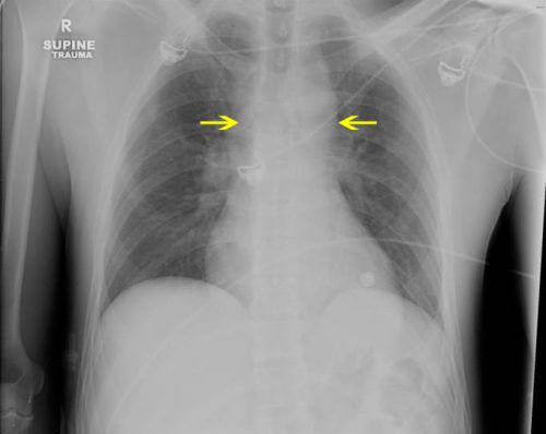
When is the last time this has happened to you? You are called to the ED for a trauma activation. The patient was involved in a motorcycle crash and is doing fine, but he has a large midline scar on his abdomen. You inquire as to what it is. He tells you that he had been involved in another motorcycle crash about five years ago and needed an operation. When questioned about what his injuries were and what was done, he has no idea.

This is an example of health (il)literacy at its best. An earlier study from the Presley trauma center in Memphis demonstrated that less than half of their trauma patients could correctly recall their injuries or their operations.

This is not really surprising. Have you ever taken a minute to look at the sheaf of paper given to hospital patients when they are discharged? They are usually computer-generated gobbledygook and are not easily understood by any human on this earth. It is hard enough to figure out the discharge medications and followup visits. And any diagnosis or surgical procedure information is never in patient-friendly language.

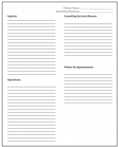
The Memphis group designed a simple discharge information form to provide to their patients:

Here are the factoids:

  • Patients admitted to the trauma service over a 6-month period were studied and surveyed during their first post-discharge clinic visit
  • A total of 153 surveys were distributed, asking about income, education, and patient satisfaction and their understanding of what happened to them; 146 were returned
  • Income levels were low, with about 60% of them less than $25K and 85% less than $50K
  • About 75% had a high school education or less
  • Implementation of the form increased injury recall some or all of patient injuries from 55% to 85%, and recall of operations from 43% to 76%
  • The number of patients who could recall any of their providers’ names increased from 11% to 31% (!)
  • Injury understanding, satisfaction with injury understanding, and the overall impact on hospitalization was significantly positive

The authors concluded that introducing this simple form dramatically improved their patients’ health literacy, and their patients were able to provide more details to providers they visited post-discharge.

Here are my comments: I think the bottom line here is to know your patients! Socioeconomic and education status vary dramatically by geographic location. This certainly has an impact on the understanding and recall of hospital events by our patients. It can help us optimize processes to provide meaningful and important information that they need to know in the future.

The form used in this study was very simple, consisting of a series of blanks to be filled in by a healthcare provider. But who was this provider? All medical professionals tend to use the lingo that we learned in training. But our patients have zero understanding of them. Consider the lowly Foley catheter. Tell a patient you are going to insert one, and they will say “uh-huh.” But tell them that you are preparing to stick a big rubber tube in their penis, and the response will be much more vocal. Make sure the language is simple and lingo-free.

The recall of provider names improved only modestly. This may be due to the typical “interchangeable head” model where the various healthcare professionals change on a frequent bases. Additionally, patients are seen by a horde of nurses, physicians, APPs, residents, techs, and others during their stay so it’s easy to forget a name.

Overall, the results were very promising. This is a significant advance in patient health education and literacy. I think the next step is to provide a library of information sheets based on the common injury diagnoses and operations that occur at the trauma center. This, coupled with a more intelligible set of discharge papers in general will be of great help to our patients.

Here are my questions for the presenter and authors:

  • Why so few surveys? Your center is very busy, and the study data only involved about 25 patients per month. How did you select them, and might information obtained from all the other patients have changed your results?
  • Did you independently review the discharge forms to ensure understandable language? The intelligibility could vary significantly based on the provider filling it out.
  • How did your care model affect the patient recall of their providers? Do your residents or attending surgeons rotate on a frequent basis? What other factors might have influenced this?
  • What next? How has this information changed how you educate your patients now? What additional changes might you make in the future? How will you roll it out to more than just 25 patients per month?

This is excellent work! I’m looking forward to your live presentation later this week.

Best Of AAST #13: Work-Life Balance

Okay, so this abstract is a bit more on the touchy-feely side. But it is extremely important because it speaks to the balancing act we all have to perform in order to achieve a satisfying harmony between work and everything else.

Older generations of surgeons threw nearly all of their energy into work, and ended up with lesser amounts of involvement with their family and everything else outside of work. At the time, , though, people seemed to be (mostly) satisfied. That’s just the way it was.

But now, there is much more emphasis on a healthy lifestyle, and this includes a healthy delineation of work and not-work. An AAST-approved survey was sent to the membership which tried to parse out the various factors involved in work-life balance, happiness, and burnout.

Here are some very interesting factoids:

  • Of more than 1300 questionnaires sent out, only 291 (21%) returned them (wish I had a sad face icon here)
  • Only 43% were satisfied with their work-life balance
  • There was no difference in satisfaction based on age, sex, or practice type
  • Here are the factors that set the satisfied surgeons apart from the dissatisfied:
    • Early (<10 years) or late in career (>20 years)
    • Fewer hours spent at work
    • More hours spent (awake) at home
    • Enjoy their job
    • Enjoy their partners
    • Better at saying no or delegating work tasks
    • Feel they are fairly compensated
    • Engage in hobbies (86% vs 68%)
    • Exercise regularly (49% vs 20%)
    • Eat a healthy diet (74$ vs 48%)
    • Get more sleep (7 hrs vs 6 hrs)
  • Despite getting the same amount of vacation time, the satisfied surgeons actually used it
  • Dissatisfied surgeons reported significantly more feelings of burnout (77% vs 39%)

The authors concluded that trauma programs should concentrate on optimizing the modifiable factors listed above to improve satisfaction and decrease burnout.

Here are my comments: Well, I don’t have many, nor do I have any questions for the authors. This is a purely descriptive study that paints a general picture outlining what seems to be important in enhancing satisfaction with one’s career path. It is an interesting read, and outlines many of the factors that influence this. I’m sure it’s not all of the factors, but they hit the big ones.

All trauma professionals should look at this data and read the final manuscript. It may help you make changes to optimize your own work-life balance and career satisfaction.

Reference: Modifiable factors to improve work-life balance for trauma surgeons. AAST 2020, Oral abstract #50.

Best of AAST #12: Embolization Of Splenic Pseudoaneurysm

The management of blunt spleen injury has evolved significant over the time I’ve been in practice. Initially, the usual formula was:

Spleen injury = splenectomy

This began to change in the late 1980’s, and beginning in the early 90’s nonoperative management became the rage. We spent the next 10-15 years tweaking the details, gradually reducing bed rest and NPO times, and increasing the success rate through smart patient selection and discovering new adjuncts.

One of these adjuncts was angiography with embolization. The ShockTrauma Center in Maryland was an early adopter and protocolized its use in patients with high-grade injuries.

But now, they are questioning the utility of this tool in certain patients: those with splenic pseudoaneurysms (PSA). They theorized that modern, high resolution CT identifies relatively unimportant pseudoaneurysms. They conducted a 5-year retrospective review of their experience.

Here are the factoids:

  • They identified 717 splenic injuries, of whom 155 were embolized but only 140 patients had adequate records and imaging for review
  • The majority of patients had high grade injury: 31% Grade 3, 61% Grade 4, 1% Grade 5
  • Extravasation was seen in 17% and PSA in 52%
  • About 44% of patients went to angiography within 6 hours, but the mean was 17 hours indicating quite a few outliers
  • Among the 73 patients with an initial PSA , a third of them did not have a detectable lesion during angiography
  • Patients who underwent embolization for PSA had a followup CT 48-72 hours afterwards, persistently perfused PSA were seen in 40% (!)
  • No patients with PSA who were only observed required delayed splenectomy

The authors conclude that a third of pseudoaneurysms may be clinically insignificant, and that 40% of them persist after embolization. They do not, however, offer any recommendations based on their data.

Here are my comments: This is an interesting study. My read of the abstract and slides would indicate that this group routinely sends all Grade 3 and 4 injuries to angio, and Grade 5 could go to either angio or OR. They take their good time going to interventional radiology (mean 17 hours from arrival), and get a routine followup CT 48-72 hours from hitting the door if they didn’t go to the OR.

If I were to play the devil’s advocate, I might think that interventional radiology was being de-emphasized for some reason. Was there some reluctance to send patients there, or limited availability? This might explain the long access times. And how are the radiologists not shutting down 40% of PSA that are seen?

I am intrigued by the study, but there are a lot more details needed to get some good takeaways from it.

Here are my questions for the presenter and authors:

  • Please explain why it takes so long to send patients to angiography. Less than half got there in less than 6 hours, and the mean of 17 hours means that many didn’t get there until the next day.
  • Does this small study have the statistical power to say that some PSA are benign? The groups are very small, and I would speculate that the group size needed to show significance is in the high hundreds.
  • What was the reason for splenectomy in the 2 patients who underwent embolization? Was it related to the pseudoaneurysm or something else?
  • How can you be sure that these PSA are insignificant? Frequently, pseudoaneurysms don’t explode for 7-10 days. Do you have any data on patients who returned to a hospital with delayed bleeding?
  • If you believe that many pseudoaneurysms are benign, how do you propose to manage the patients? Observe until they explode? Repeat a contrast CT scan, with the associated contrast and radiation re-dose? And how long would you wait to do this? What would your new protocol be?

I’ll be all ears on Friday when this abstract is presented live.