All posts by The Trauma Pro

VIP Syndrome In Healthcare (Very Important Person)

Current events are making this classic post even more poignant!

The VIP syndrome occurs in healthcare when a celebrity or other well-connected “important” person receives a level of care that the average person does not. This situation was first documented in a paper published in the 1960s which noted that VIP patients have worse outcomes.

Who is a VIP? It may be a celebrity. A family member. Or even a colleague. Or the President of the United States. VIPs (or their healthcare providers) may have the expectation that they can get special access to care and that the care will be of higher quality than that provided to others. Healthcare providers often grant this extra access, in the form of returned phone calls and preferential access to their clinic or office. The provider tries to provide a higher quality of care by ordering additional tests and involving more consultants. This idea ignores the fact that we already provide the best care we know how, and money or fame can’t buy any better.

Unfortunately, trying to provide better care sets up the VIP for a higher complication rate and a greater chance of death. Healthcare consists of a number of intertwined systems that, in general, have found their most efficient processes and lowest complication rates. Any disturbance in this equilibrium of tests, consultants, or nursing care moves this equilibrium away from its safety point.

Every test has its own set of possible complications. Each consultant feels compelled to add something to the evaluation, which usually means even more tests, and more possible complications. And once too many consultants are involved, there is no “captain of the ship” and care can become fragmented and even more inefficient and dangerous.

How do we avoid the VIP Syndrome? First, explain these facts to the VIP, making sure to impress upon them that requesting or receiving care that is “different” may be dangerous to their health. Explain the same things to allproviders who will be involved in their care. Finally, do not stray from the way you “normally” do things. Order the same tests you usually would, use the same consultants, and take control of all of their recommendations, trying to do things in your usual way. This will provide the VIP with the best care possible, which is actually the same as what everybody else gets.

Reference: “The VIP Syndrome”: A Clinical Study in Hospital Psychiatry. Weintraub, Journal of Mental and Nervious Disease, 138(2): 181-193, 1964.

Does Time To Interventional Radiography Make a Difference In Solid Organ Injury?

Solid organ injury is one of the more common manifestations of blunt abdominal trauma. Most trauma centers have some sort of practice guideline for managing these injuries. Frequently, interventional radiology (IR) and angioembolization (AE) are part of this algorithm, especially when active bleeding is noted on CT scan.

So it makes sense that getting to IR in a timely manner would serve to stop the bleeding sooner and help the patient. But in most hospitals, interventional radiology is not in-house 24/7. Calls after hours require mobilization of a call team, which may be costly and take time.

For this reason, it is important to know if rapid access to angioembolization makes sense. Couldn’t the patient just wait until the start of business the next morning when the IR team normally arrives?

The group at the University of Arizona at Tucson tackled this problem. They performed a 4-year retrospective review of the TQIP database. They included all adult patients who underwent AE within four hours of admission. Outcome measures were 24-hour mortality, blood product usage, and in-hospital mortality.

Here are the factoids:

  • Out of over a million records in the database, only 924 met the inclusion criteria
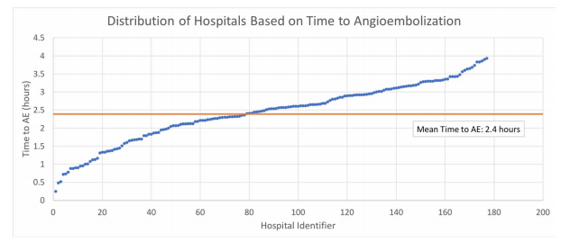
  • Mean time to AE was 2 hours and 22 minutes, with 92% of patients getting this procedure more than an hour after arrival
  • Average 24-hour mortality was 5%. Mortality by hours to AE was as follows:
    • Within 1 hour: 2.6%
    • Within 2 hours: 3.6%
    • Within 3 hours: 4.0%
    • Within 4 hours: 8.8%
  • There was no difference in the use of blood products

The authors concluded that delayed angioembolization for solid organ injury is associated with increased mortality but no increase in blood product usage. They recommend that improving time to AE is a worthy performance improvement project.

Bottom line: This study has the usual limitations of a retrospective database review. But it is really the only way to obtain the range of data needed for the analysis. 

The results seem straightforward: early angioembolization saves lives. What puzzles me is that these patients should be bleeding from their solid organ injury. Yet longer delays did not result in the use of more blood products.

There are two possibilities for this: there are other important factors that were not accounted for, or the sample size was too small to identify a difference. As we know, there are huge variations in how clinicians choose to administer blood products. This could easily account for the apparent similarities between products given at various time intervals to AE.

My advice? Act like your patient is bleeding to death. If the CT scan indicates that they have active extravasation, they actually are. If a parenchymal pseudoaneurysm is present, they are about to. So call in your IR team immediately! Minutes count!

Reference: Angioembolization in intra-abdominal solid organ injury:
Does delay in angioembolization affect outcomes?  J Trauma 89(4):723-729, 2020.

In The Next Trauma MedEd Newsletter: Fatigue And Sleep Loss

The next issue of Trauma MedEd will be sent out to subscribers this week, and will provide some interesting information on fatigue and sleep loss.

Most trauma professionals have at least a little experience with this topic. You may one who provides care on call at night, or work a crazy shift system. We all end up on the short end of the sleep stick at times.

Topics will include:

  • Facts on fatigue and sleep loss
  • Impact on EMS providers
  • Impact of Nurses
  • Impact on Physicians and APPs
  • What to do about it

As always, this month’s issue will go to all of my subscribers first. If you are not yet one of them, click this link right away to sign up now and/or download back issues.

Unfortunately, non-subscribers will have to wait until I release the issue on this blog, in mid-June. So sign up now!

How To Detect Bucket-handle Intestinal Injuries With CT Scan

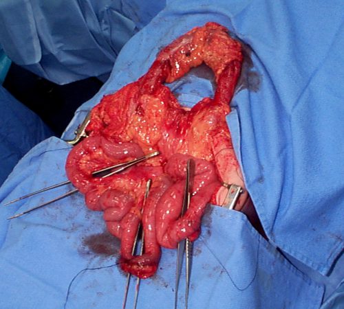
A bucket-handle injury is a relatively uncommon complication of blunt trauma to the abdomen. It only occurs in a few percent of patients, but is much more likely if they have a seat belt sign.  The basic pathology is that the bowel mesentery (small bowel of sigmoid colon) gets pulled away from the intestinal wall.

This injury is problematic because it may take a few days for the bowel itself to die and perforate. Patients with no other injuries could potentially be discharged from the hospital before they become overtly symptomatic, leading to delayed treatment.

Here’s an image from my personal collection with not one, but four bucket-handle injuries.

Typical patients with suspected blunt intestinal injury are observed with good serial exams and a daily WBC count. If this begins to rise after 24 hours, there is a reasonable chance that this injury is present.

CT scan has not really been that reliable in past studies. There may be some “dirty mesentery”, which is contused and has a hematoma within it. But without a more convincing exam, it is difficult to convince yourself to operate immediately on these patients.

A paper was published by a group of radiologists at Duke University. It appears to be a case report disguised as a descriptive paper. It looks like they picked a few known bucket-handle injuries from their institution and back-correlated them with CT findings.

The authors called out the usual culprits:

  • Fluid between loops of bowel
  • Active bleeding in the mesentery
  • Bowel wall perfusion defects

But they also noted that traumatic abdominal wall hernias were highly with the injury as well. These are rare, but should bring intestinal injury to mind when seen.

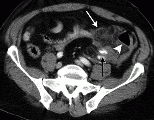
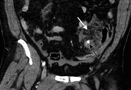
With newer scanners, radiologists are better able to detect subtle areas of hypoperfusion as well. This is a fairly good indicator of injury, especially when adjacent bowel appears normally perfused. Here are two examples. The black arrows denote active extravasation, and the white ones an area of hypoperfusion.

The authors add bowel wall hypoperfusion as another finding that may point to a bucket-handle type injury

Bottom line: Hold the phone! Don’t change your practice yet. This paper is not able to demonstrate how good this radiographic sign is. Looking at other radiology literature, the specificity is about 90%. But remember, that means that if they don’t have the CT finding, that’s true only 90% of the time.

Unfortunately the sensitivity is only 10%. So if you see it on the scan, they’ve got a 1 in 10 chance of actually having the injury. That’s not good enough for me to run to the operating room.

Here’s what I recommend: if your patient has an unconcerning exam and any of the usual culprits (pelvic fluid, inter-loop fluid, dirty mesentery, thickened bowel loops, abdominal wall hernia), perform serial exams and get a WBC the next morning. If the exam worsens, operate. If the WBC rises, consider laparoscopy to see if you need to make a bigger incision. And if you see this new kid on the block, the hypoperfused bowel, consider laparoscopy right away. 

I’m sure the radiologists and the technology will keep getting better. But for now, blunt intestinal injury still requires patience, perceptiveness, and a little luck.

References:

  • CT findings of traumatic bucket-handle mesenteric injuries. Am J Radiol 209:W360-@364, 2017.
  • Multidetector CT of blunt abdominal trauma. Radiology 265(3):678–693, 2012.

What Is A Wide Mediastinum Anyway?

Trauma professionals are always on the lookout for injuries that can kill you. Thoracic aortic injury from blunt trauma is one of those injuries. Thankfully, it is uncommon, but it can certainly be deadly.

One of the screening tests used to detect aortic injury is the old-fashioned chest xray. This test is said to be about 50% sensitive, with a negative predictive value of about 80%. However, the sensitivity is probably decreasing and the negative predictive value increasing due to the rapidly increasing number of obese patients that we see.

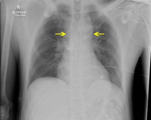
A wide mediastinum is defined as being > 8cm in width. In this day and age of digital imaging, you will need to use the measurement tool on your workstation to figure this out.

Unfortunately, it seems like most chest xrays show wide mediastinum these days. What are the most common causes for this?

  • Technique. The standard xray technique used to reduce magnification of the anterior mediastinum (where the aortic arch lives) is a tube distance of 72 inches from the patient, shot back to front. We can’t do this for trauma patients because we can’t stand them up and are reluctant to prone them. The standard trauma room technique is 36 inches from the patient shot front to back. This serves to magnify the mediastinal image and make it look wide.
  • Obesity. The more fat in the mediastinum, the wider it looks. The more fat on the back, the further the mediastinum is from the xray plate and the greater the magnification.
  • Other mediastinal blood. Major blunt trauma to the chest can cause bleeding from small veins in the mediastinum, making it look wide.
  • Thymus. Only in kids, though.
  • Aortic injury. Last but not least. Only a few percent of people with wide mediastinum will actually have the injury.

If you encounter a wide mediastinum on chest xray in a patient with a significant mechanism for aortic injury, then they should be screened using helical CT.