All posts by The Trauma Pro

EAST Guidelines: TEG And ROTEM In Coagulopathic Trauma Patients

In my last post, I explained why TEG is not so easy to use. Today, I’ll share the new Eastern Association for the Surgery of Trauma (EAST) practice management guidelines for using TEG and its twin, ROTEM for bleeding patients.

TEG first appeared in the trauma literature in 2008. A paper by John Holcomb showed that it was superior to the standard lab tests (PT, aPTT, and activated clotting time) in monitoring hemorrhagic shock in pigs. Since then, research has exploded with TEG papers. There have been about 50 published annually for the last four years.

In this month’s Journal of Trauma, EAST published their most recent practice management guideline, dedicating it to TEG. They identified over 6,000 potential papers and ultimately settled on 38 articles. They used them to attempt to answer three questions regarding use of these devices during resuscitation.

Question 1

In adult trauma patients with ongoing hemorrhage, should TEG/ROTEM be used vs non-TEG/ROTEM monitoring to guide transfusion strategy in order to reduce mortality, blood product transfusions and the need for additional hemostatic interventions such as angioembolization, endoscopy, or operation?

Answer: Only seven studies were found regarding this question. All but one showed no difference in 24 hour and hospital mortality. They also showed an inconsistent effect on blood product usage with some showing no difference and some shower less transfused product.

Nonetheless, EAST “conditionally recommended” the use of TEG/ROTEM. This is based  solely on the presumption that it can reduce the risk of blood transfusions by using a test that is harmless.

Question 2

In adult surgery patients with ongoing hemorrhage, should TEG/ROTEM be used vs non-TEG/ROTEM monitoring to guide transfusion strategy in order to reduce mortality, blood product transfusions and the need for additional hemostatic interventions such as angioembolization, endoscopy, or operation? Note the shift here to non-trauma patients.

Twenty one studies were found addressing this question. Most papers showed no difference in reoperation rate. There were also no consistent differences in transfusion of various blood products. And the vast majority showed no difference in mortality.

But once again, EAST conditionally recommended the use of this test in these patients, mainly because it is believed to be harmless.

Question 3

In adult critically ill patients with ongoing hemorrhage, should TEG/ROTEM be used vs non-TEG/ROTEM monitoring to guide transfusion strategy in order to reduce mortality, blood product transfusions and the need for additional hemostatic interventions such as angioembolization, endoscopy, or operation?

There were only 10 studies relating to this question, and they included patients with a variety of surgical and medical problems. TEG/ROTEM was no better than non-TEG parameters in predicting the need to transfuse, but did somewhat better than clinical judgement. Once again, there was no consistent effect on the number of transfusions given, although some studies showed that use of non-TEG/ROTEM studies resulted in fewer units of red cells, platelets, and cryoprecipitate given.

Interestingly, although there was little difference in the number of units transfused, fewer patients required transfusion using TEG/ROTEM. There was no difference in mortality or interventions to stop bleeding.

Yet again, EAST conditionally recommended use of TEG/ROTEM in these patients despite the very low level of evidence. Again, this is mainly because of the lack of perceived harm in using it, and the possibility that it might reduce exposure to blood products.

Bottom line: Hmm. I remain skeptical. What EAST is saying is that, hey it’s harmless and there’s a chance that it might reduce a patient’s exposure to blood products, so why not? I have a vial of bat wings and eye of newt that might do the same thing. As long as it’s harmless, right?

Well, it may be clinically harmless, but it costs money and time. First, you have to buy the machine. Luckily, they are much cheaper than a CT scanner. But then the manufacturer kills you with the disposables. Like a cheap inkjet printer, you have to keep buying $40 ink cartridges every few weeks to keep it working. Except TEG cartridges cost more than $40.

And don’t overlook the time spent training people in how to interpret the curves. And developing a system to obtain the specimen and pay people to run the equipment. It all adds up, and yet the papers can’t show us any dramatic clinical results.

I’ll probably irritate the TEG/ROTEM true believers, but it still seems like a device searching for a great clinical problem to solve. IMHO we need much more high-quality research to help us figure out how this tool can help us with our trauma / surgical / critical care patients.

Reference: Thromboelastography and rotational thromboelastometry in bleeding patients with coagulopathy: Practice management guideline from the Eastern Association for the Surgery of Trauma. J Trauma 89(6):999-1017, 2020.

How To Make TEG / ROTEM Useful

A lot of papers have been written on the use of thromboelastography in trauma. And pretty much any meeting or course you may attend has at least one talk on it. And I get it. It can be an important tool in treating trauma patients who have some sort of coagulation disturbance. It helps us figure out what specific part of the coagulation process is out of whack and suggests how we can fix it.

But there are a few problems. And the “friction” that those issues cause overall decreases how useful it is. Here’s a partial list of the problems:

  • The equipment costs money, and the disposables that must be used for every patient do, too.
  • Where do you put the machine? Most hospitals can’t put one unit in every possible area it might be used.
  • How to you get the results to a care area if there is no unit there?
  • There is a significant learning curve for interpreting the results
  • How can it be integrated into the massive transfusion protocol?

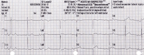
The main issue is that the current state of TEG and ROTEM are very similar to the state of electrocardiography shortly after it’s discovery. Here’s what you got then:

In order to get the most from an EKG, you need to combine this tracing with that from other leads, do a bunch of measurements, look for abnormal shapes and elevations/depressions, etc.  This is exactly where we are with TEG and ROTEM today. Relatively crude, and it takes a lot of work to use it.

The tracing below shows where we are with EKGs today. A computer program looks at all the tracings, and rapidly applies a complex set of rules to come to a set of diagnoses. Notice in the image below that this reading is “unconfirmed.” But how many times in your career have you seen a cardiologist correct one of these? The machines are actually very good!

Bottom line: The tracing above is where we need to be with TEG and ROTEM. Instead of a clinician staring at a developing tracing and figuring out what products to give, these machines need to be just like an automated EKG machine. Sure, a human can still stare at the trace. But the machine will automatically monitor it, apply rules about what abnormalities are present and what is needed to correct them. Send off your blood specimen, and within minutes instructions like “infuse 2 units of plasma now” or “give 12u cryo now” appear. These may be displayed on a monitor in the treatment area, or be broadcast to the phone or pager of the responsible clinicians.

Current TEG/ROTEM equipment is what I would consider 1st generation. The next generation will reduce or remove much of the “friction” in the current process and allow us to really integrate TEG/ROTEM meaningfully into the massive transfusion protocol for trauma. And for anyone who develops this 2nd generation equipment, don’t forget my royalty checks for this idea! 

In my next post, I’ll review the new EAST guidelines for the use of TEG and ROTEM.

Should I Apply Compression Devices To Patients With DVT?

Everyone knows that venous thromboembolism (VTE) is a potential problem in hospitalized patients, and especially so in trauma patients. Several groups of them are at higher risk by virtue of the particular injuries they have sustained and the activity restriction caused.

Nearly every trauma program uses some form of screening and prophylaxis in an attempt to reduce the occurrence of this problem, which can result in deep venous thrombosis (DVT) and/or pulmonary embolism (PE). Screening looks at patient factors such as age, obesity, previous VTE as well as injury risk factors like spine and pelvic fractures, and decreased mobility.

Based on the screening protocol, prophylaxis may be prescribed depending upon level of VTE risk, which is then balanced with bleeding risk from brain, solid organ, or other injuries. The choices we have are primarily mechanical vs chemical and consist of compression devices (sequential or not) and various heparins.

An age old question surfaced on my own patient rounds recently. If a patient breaks through their prophylaxis and develops DVT, is it safe to apply compression devices to the extremity?

There has always been the fear that doing things that increase flow in the affected extremity may cause clots to dislodge and ultimately cause a PE. Seems logical right? But we know that often, our common sense about things is completely wrong.  Couldn’t just moving around cause pieces to break off? A meta-analysis of 13 studies published in 2015 showed that early ambulation was not associated with a higher incidence of new PE. Furthermore, patients who suffered from pain in the affected extremity noted significant improvements with early ambulation.

If ambulation makes the pain better, could the veins be recanalizing more quickly? Another study examined a small group of 72 people with DVT receiving anticoagulants, half of whom were prescribed exercise and compression stockings and the other half stockings only. There was a huge amount of variability in the rates of recanalization, but ultimately there were no significant differences with or without exercise.

So just lying in bed is not good, and exercise/ambulation may actually make people feel better. But interestingly, bedrest alone does not appear to increase the likelihood of PE! It does decrease the risk of developing problems other than the VTE, like pulmonary complications.

But what about compression devices? Common sense would say that you are intermittently  increasing pressures in the leg veins, which could dislodge any loose clots and send them flying to the lungs, right?

Unfortunately, I couldn’t find a paper from anyone who had the courage to try this. Or perhaps no institutional review board (IRB) would approve it. But the key fact is that every compression device manufacturer includes existing DVT as a contraindication in their product documentation. They don’t have any literature either, so I assume it’s an attempt to limit litigation, just in case.

Bottom line: Walking provides at least as much muscle compression as compression devices. But the simple truth is that we have no solid research that either supports or condemns the use of active compression devices in patients with known DVT. And we probably won’t, ever.

Compression stockings seem to be safe, but they really don’t do much. They are white, but don’t do much more than contribute to hospital clothing fashion. Since the manufacturers define existing DVT as a contraindication, application of their product would be considered an off-label use. So it looks like we cannot in good faith use these devices in patients with diagnosed DVT.

References:

  • Bed Rest versus Early Ambulation with Standard Anticoagulation in The Management of Deep Vein Thrombosis: A Meta-Analysis. PLOS One , April 10, 2015, https://doi.org/10.1371/journal.pone.0121388
  • Bed Rest or Ambulation in the Initial Treatment of Patients With Acute Deep Vein Thrombosis or Pulmonary Embolism: Findings From the RIETE Registry. Chest 127(5):1631-1636, 2005.
  • Does supervised exercise after deep venous thrombosis improve recanalization of occluded vein segments? A randomized study. J Thrombosis Thrombolysis 23:25-30, 2006.

Delayed Presentation Of Right Diaphragm Injury

Diaphragm injury from blunt trauma is uncommon, occurring in only a few percent of patients after high energy mechanisms. They usually occur on the left side, and are more frequently seen after t-bone type car crashes and in pedestrians struck by a car.

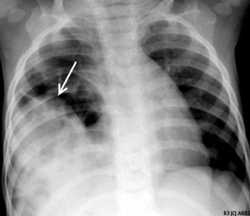
Blunt diaphragm injury on the right side is very unusual. Even so, it is more easily detected due to obvious displacement of the liver that can be seen on chest x-ray. Blunt injuries on the right side usually result in a large rent in the central tendon, or detachment of the diaphragm from the chest wall. This allows the liver to herniate into the chest, and the chest x-ray finding is not subtle.

This image shows an acute herniation of the liver through the diaphragm. Due to the size of the liver, only part of it can typically fit through the rent. Radiologists call this the “cottage loaf” sign. Why? Here’s the bakery item it is named after. Get it now?

Thankfully, most of these injuries are identified in the acute setting. They must be addressed surgically because, if left untreated, more and more of the liver will slowly move into the chest resulting in respiratory problems in the long run.

Acute management usually consists of laparotomy to address both the diaphragm tear and any other associated intra-abdominal injuries. The liver should be reduced by sliding a hand next to it laterally into the chest cavity and pushing the dome downwards. The right triangular ligaments should be taken down (if they are not already destroyed) to mobilize the organ better so the diaphragm laceration can be closed. This is typically accomplished with some type of large (size 0) permanent suture. A chest tube will be needed to evacuate the iatrogenic pneumothorax created by opening the abdomen.

Chronic right diaphragm injuries are a different animal entirely. There is no longer any need to evaluate for intra-abdominal injury, so the procedure is usually performed through the chest. For smaller injuries, thoracoscopic procedures have been described that push the liver downwards and then either suture the diaphragm primarily or (more likely) incorporate a piece of mesh.

Larger injury requires conversion to an open procedure so more muscle power can be used to push the liver downwards to facilitate the repair. However, do not underestimate the adhesions that will be present between diaphragm and liver (and possibly the lung) in long-standing injuries. It may take some time to dissect them away. Rarely, a laparotomy (or laparoscopy) may be needed to assist for very large and complex injuries.

References:

  • Management of Delayed Presentation of a Right-Side Traumatic
    Diaphragmatic Rupture. World J Surg 36:260-265, 2012.
  • Delayed Discovery of Diaphragmatic Injury After Blunt Trauma:
    Report of Three Cases. Surg Today 35:407-410, 2005.

Does Time To Angioembolization Make A Difference?

Angiography and angioembolization (AE) have helped trauma professionals stop pesky bleeding from the pelvis, solid organs, and other hard to reach places for decades. The American College of Surgeons has recognized its importance and even has a specific criterion for interventional radiologist response times.

Many centers have found this 30-minute response for radiologist arrival to be onerous. So, of course, someone decided to look at the data to see if it was really warranted. The group at the University of Arizona at Tucson did a big data dive using the Trauma Quality Improvement Program database to try to tease out the consequences of prompt vs delayed access to AE in blunt trauma patients.

Four years of TQIP data were analyzed. The authors focused on the records of patients who underwent AE within 4 hours of arrival for blunt injury to liver, spleen, or kidney. They excluded transfers in, burn patients, and those who underwent an operation prior to AE. The 4 hour AE period was subdivided into hourly segments and outcomes were examined for each.

Here are the factoids:

  • Out of over a million records, 924 met study inclusion criteria
  • Patients were relatively young with a mean age of 44, and seriously injured with a median ISS of 29
  • Spleen injuries were most common (64%), followed by liver (50%), and kidney (27%)
  • Overall 24 hour mortality was 5% and in-hospital mortality was 15%
  • The 24 hour mortality significantly increased as each hour passed until AE, from 2% to 24%. 
  • On multivariate analysis, in-hospital mortality showed the same hourly increase
  • No differences in the amount of blood, plasma, or platelets given were noted in any of the groups
  • Average time to AE correlated with trauma center AE volume, with 1.6 hours at centers doing more than 14 cases per year to 2.7 hours at those doing less than 9 cases

The authors concluded that delayed AE for solid organ injury resulted in increased mortality without any difference in transfused blood products. They encouraged centers to ensure rapid access to this vital procedure.

Bottom line: This is an important paper. Other research on angiography in blunt trauma has not shown the survival differences that we see here. But they did not focus on patients who underwent actual embolization. This one shows that time to AE is very important in those patients who really need it.

It appears that the most important factor is door to angiography suite time. There are many factors involved, though. First, the specific injury must be recognized by the clinicians. This involves access to CT, timeliness of radiologist report, decision making by the trauma professional, and timeliness of response by the interventional radiologist and the IR team.

Most centers focus only on that 30-minute response time required of the interventionalist. But as you can see, there are many more moving parts. I would urge all centers to look at their door to AE time and compare to what was found in this study. If you find that it is taking more than an hour and a half, it’s probably time for a PI project to tighten it up. This is particularly important in centers with low AE volume.

Reference: Angioembolization in intra-abdominal solid organ injury: Does delay in angioembolization affect outcomes? J Trauma 89(4):723-729, 2020.