All posts by The Trauma Pro

Arms Up or Arms Down In Torso CT Scans?

CT scan is a valuable tool for initial screening and diagnosis of trauma patients. However, more attention is being paid to radiation exposure and dosing. Besides selecting patients carefully and striving for ALARA radiation dosing (as low as reasonably achievable) by adjusting technique, what else can be done? Obviously, shielding parts of the body that do not need imaging is simple and effective. But what about simply changing body position?

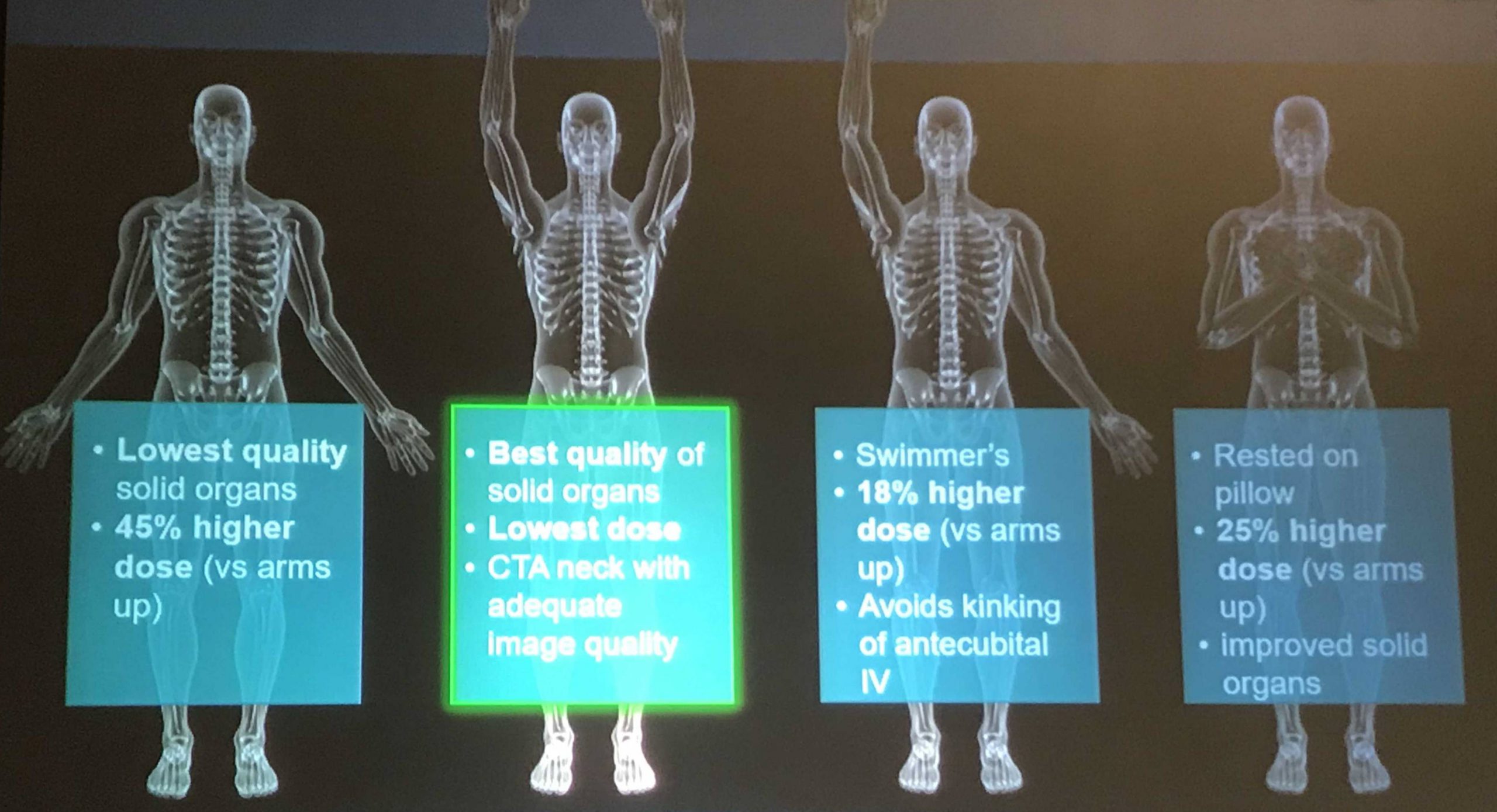
One simple item to consider is arm positioning in torso scanning. There are no consistent recommendations for use in trauma scanning. Patients with arm and shoulder injuries generally keep the affected upper extremity at their side. Radiologists prefer to have the arms up if possible to reduce scatter and provide clearer imaging.

Radiation physics research has examined arm positioning and its effect on radiation dose. A retrospective review of 690 patients used dose information computed by the CT software and displayed on the console. Radiation exposure was estimated using this data and was stratified by arm positioning. Even though there are some issues with study design, the results were impressive.

The dose results were as follows:

  • Both arms up: 19.2 mSv (p<0.0000001)
  • Left arm up: 22.5 mSv
  • Right arm up: 23.5 mSv
  • Arms down: 24.7 mSv

Bottom line: Do everything you can to reduce radiation exposure:

  1. Be selective with your imaging. Do you really need it?
  2. Work with your radiologists and physicists to use techniques that reduce dose yet retain image quality
  3. Shield everything that’s not being imaged.
  4. Think hard about getting CT scans in children
  5. Raise both arms up during torso scanning unless injuries preclude it.

Reference: Influence of arm positioning on radiation dose for whole body computed tomography in trauma patients. J Trauma 70(4):900-905, 2011.

Antihypertensive Treatment In Acute TBI

Yes, we know high blood pressure can be bad. Over the long term, it can accelerate atherosclerotic heart disease and pound away at the kidneys and brain. And when it is acutely elevated to critical levels, it can lead to stroke.

But is it always bad in trauma? Trauma hurts like hell, so it’s no wonder than many of our patients (not suffering blood loss of course) are hypertensive.  But how often have you seen this scenario occur:

An elderly patient fell from standing, striking her head. She is brought to your ED by ground EMS. She has a GCS of 8 (E1 V3 M4) with a BP of 200/130 and pulse of 56.  This meets your trauma activation criteria and the team assembles to meet the patient.

As you move her onto the bed, one of your colleagues calls out for some nicardipine to control the pressure. Is this a wise move? Remember the First Law of Trauma:

Any anomaly in your trauma patient is due to trauma, no matter how unlikely it may seem.

What else can cause hypertension and bradycardia in your trauma patient? In this case, certainly a subdural or epidural hematoma.

And why is that happening? Because the intracranial pressure is elevated from the space-occupying lesion. Remember the formula for cerebral perfusion pressure (CPP):

CPP = MAP – ICP

Where MAP = mean arterial pressure and ICP = intracranial pressure.  Normally the MAP is around 90 torr and ICP is about 10 torr. Thus, the normal CPP is approximately 80. The range is 60  to well over 100 torr, and flow autoregulation keeps brain perfusion constant over this range.

But let’s say that we are psychic and know the ICP of our patient to be 60 because of a large subdural hematoma. Her current CPP is 150 – 60 or about 90 torr. What happens if we start her on a nicardipine drip or some other antihypertensive medication? We can certainly normalize the blood pressure to 120/80. But now her CPP drops to 90  – 60 = 30 torr!

Congratulations, you have just shut down circulation to her brain!

Bottom line: Think first before calling for antihypertensive medications in patients who may have increased intracranial pressure. You may be sabotaging the only mechanism protecting their brain while you are calling your neurosurgeon for help. Your top priority is to get them to the CT scanner while permitting that pressure. If it turns out that there is no evidence for pathology that would lead to increased ICP, then turn to the antihypertensive agents to help protect against stroke. 

Using Chest CT To Detect Occult Injuries

There are major belief systems when it comes to the use of trauma CT: selective scan vs pan scan. The selective scanners believe that too much radiation can be bad, and that the risk of excess exposure outweighs the value of scanning everything. The pan scanners believe that valuable information might be missed unless they routinely image everything.

Who is right? There’s probably value in each side of the argument. But do we have data? Good data? Two emergency medicine groups from UC-Irvine and UC-San Diego tried to answer this question via a prospective study involving 10 Level I trauma center EDs in California.  They tagged onto data collection underway for the NEXUS chest and chest CT studies from 2009-2012.

Patients with fresh (< 24 hours) blunt trauma who underwent chest imaging in the ED were included. Patients needed to have both CT scan and chest x-ray within 24 hours, at the discretion of the emergency physician. Weirdly, they skewed their sample by enrolling patients from 7am to 11pm daily due to availability of research personnel.

The researchers were looking for minor and major interventions necessitated by data discovered on the CT scan. Occult injuries were defined as clinically important if an intervention occurred because of it. Major interventions included surgery, mechanical ventilation for pulmonary contusion, or chest tube for hemo- and pneumothoraces.

Here are the factoids:

  • Nearly six thousand patients were enrolled, and 2,048 had at least one injury identified on either study
  • A total of 1,454 of these injuries (71% of injuries, but only 25% of patients) were occult, only being seen on the CT scan
  • Chest x-ray found all injuries in only 29% of patients (not surprising)
  • When pulmonary contusion was seen by CT only, 6% were placed on ventilators; when hemo- or pneumothorax were seen, 41% and 29% respectively had chest tubes inserted (wow!)
  • The authors tallied 241 major interventions for occult injury in 202 patients, 154 chest tubes for hemo/pneumothorax and/or mechanical ventilation, 9 operations for diaphragm or aortic injury, and the remainder appear to be for other chest wall fractures

The authors concluded that occult injuries were found in 71% of their patients, with the majority of those “requiring” chest tubes. They recognized some of the shortcomings in their study and stopped short of recommending a pan-scan type approach to major chest trauma.

Bottom line: This argument always boils down to diagnostic yield vs money vs radiation. Radiologists like to find as many things as they can, so CT is great. For me, it always comes back to that old saying: “if a tree falls in the woods when no one is around, does it make a sound?” 

The corollary is “if a diagnosis is found on CT that is not clinically relevant, do we care?” But wait, you say, they did have to intervene. Or did they?

Have you ever scanned a chest and seen something that makes you intubate the patient immediately and put them on a ventilator? Probably not. It’s a clinical judgement. The scan may make you a bit more wary, but you will still wait for some clinical signs that the patient needs that extra help. 

And what about chest tube insertion? I’m sure most of you have seen a modest pneumothorax on chest x-ray (1 cm away from the chest wall, extending to the 6th intercostal space, say). Ho hum. And then you get a CT scan and your eyes widen. It always looks much larger on the scan. It always does. Yet the patient is still lying there comfortably, with normal oxygen saturations. Do you really need to put a tube in? For decades, we used only the x-ray, and patients did fine.

So I don’t buy that the CT result made them do the interventions. It was the clinician’s choice based on how they interpreted the scan, not the patient’s clinical condition. Without specific guidelines that determine when interventions are indicated, it just boils down to “I do an intervention when I think the patient needs it.” And every clinician will have their own criteria and thresholds. It’s tough to learn from things done this way.

So I stick by my guns. We know that chest x-ray is flawed. But it does provide good clinical data even without a bunch of diagnostic minutiae. A good practice guideline that helps select the patients most likely to benefit from a CT scan is paramount.

As you can probably tell, I’m a selective scan kind of guy and still have not run across a study that is clean and compelling enough to make me change. And I think I’ll be waiting for a while for one of those to pop up!

Reference: Prevalence and clinical import of thoracic injury identified by chest computed tomography but not chest radiography in blunt trauma: multicenter prospective cohort study. Ann Emerg Med 66(6):589-600, 2015.

Is Intubation For Low GCS Necessary? Dangerous?

More dogma? I was taught that as the Glasgow Coma Scale (GCS) score drops toward 8, the higher the consideration of intubating the patient. And that a GCS 8 was pretty much an absolute indication for inserting the endotracheal tube. The rationale was that the more obtunded the patient was, the less able they were to protect their airway.

Even ATLS, our trauma textbooks, and practice guidelines from the likes of EAST recommend intubation for GCS 8 and less.

Having said that, I know many of you have been in a situation where you have a patient with GCS 8 or so, and they are lying there breathing peacefully with good oxygenation and ventilation. Do you really need to put in that tube? And we also tend to be very forgiving with obtunded children, avoiding premature intubation there as well.

Intubation is not a benign procedure. There is the potential for mayhem during the process, ICU admission will be required, and a host of ventilator and sedation-related complications are possible once the patient arrives there.

The trauma group at LAC + USC decided to look into this. They performed a five-year retrospective study of data from the TQIP database. A subset of patients was selected with isolated blunt head injury and GCS 7-8 who did not require immediate operation upon arrival. They were divided into intubation and non-intubation groups, and these were further subdivided into intubation within an hour of arrival, intubation after an hour, and never intubated groups.

Here are the factoids:

  • A total of 2,727 patients were studied; about two thirds were intubated within an hour, a quarter were never intubated, and the remaining 9% were intubated after the first hour
  • Immediately intubated patients were significantly younger and had fewer comorbidities
  • Mortality was 19% in the immediate intubation group vs 27% in the delayed group vs 11% in the never intubated group
  • Complications were significantly higher after immediate intubation, particularly DVT and ventilator associated pneumonia (VAP)
  • Regression analysis indicated that immediate intubation was independently associated with mortality compared to late or never intubated patients
  • Using additional regression testing, the authors concluded that the following two subsets of patients would benefit most from early intubation:
    • Younger patients (age < 45) with head AIS 5
    • Patients age <65 with head AIS 5

The authors recommend that “future research focus on more adequate parameters to identify patients requiring immediate intubation and should avoid an isolated fixed GCS threshold.”

Bottom line: This is a difficult paper to understand (at least for me). It looks like the authors are saying we should avoid immediate intubation of severe TBI patients with depressed GCS to reduce mortality and complications.

But you need to read the whole paper closely to really get it. First, let’s look at those mortality figures. The mortality in the three groups was:

  • intubated < 1 hour after arrival – 18.7% (from n = 1,866)
  • intubated > 1 hour after arrival – 27.4% (from n = 223)
  • never intubated – 11.4% (from n = 638)
  • If you combine the last two lines you get the mortality in the non-immediate intubations = 15.5% (from n = 861)

The authors then claim that the mortality for immediate intubation is greater than non-immediate intubation (the other two groups). This may be somewhat misleading, because the delayed intubation group actually has a higher mortality than the immediate group (27%)! This fact is covered up by combining delayed intubation with the never intubated group, bringing the number down to 15.5%!! Why shouldn’t you say that intubating the patient at any time is bad, immediate or delayed??

They suggest some criteria to try to focus in on the patients who really need intubation: younger patients (age < 45 or < 65) with head AIS 5 and GCS 7. Unfortunately, you can’t determine which patients have an AIS 5 in their head without a head CT, which may push them into the higher mortality delayed intubation group.

Remember, this type of study can only show an association, not cause and effect. The authors suggest that early intubation results in more deaths and complications. My suspicion is that patients with severe TBI don’t do poorly because they were intubated. I believe that they were intubated because the clinicians feared that they would do poorly. Unfortunately, this is information that can only be gleaned from a prospective study, not a retrospective database review.  And no amount of statistical manipulation or regression analysis can make up for this shortcoming.

This is yet another one of those studies that ends by concluding that a better study should be done. That would be okay if this one actually provided a hint that the endeavor would be worthwhile. But it doesn’t. I didn’t really learn anything from it, unfortunately.

So I still heartily recommend using your existing training, guidelines, and judgement to intubate these patients early and safely!

Reference: Isolated traumatic brain injury: Routine intubation for GCS 7 or 8 may be harmful! J Trauma, publish ahead of print, DOI: 10.1097/TA.0000000000003123, Feb 16, 2021.
The Lowly Blood Pressure Cuff: Is It Accurate?

Yesterday, I described how the typical automated oscillometric blood pressure cuff works. We rely on this workhorse piece of equipment for nearly all pressure determinations outside of the intensive care unit. So the obvious question is, “is it accurate?”

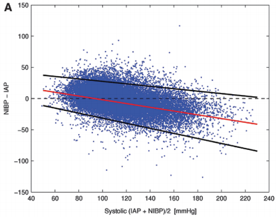
Interestingly, there are not very many good papers that have ever looked at this! However, this simple question was addressed by a group at Harvard back in 2013. This study utilized an extensive ICU database from 7 ICUs at the Beth Israel Deaconess Medical Center. Seven years of data were analyzed, including minute by minute blood pressure readings in patients with both automated cuffs and indwelling arterial lines. Arterial line pressures were considered to be the “gold standard.”

Here are the factoids:

  • Over 27,000 pairs of simultaneously recorded cuff and arterial line measurements from 852 patients were analyzed
  • The cuff underestimated art line SBP for pressures at or above 95 torr
  • The cuff overestimated SBO for pressures below 95 torr (!)
  • Patients in profound shock (SBP < 60) had a cuff reading 10 torr higher
  • Mean arterial pressure was reasonably accurate in hypotensive patients

sbp-cuff-v-aline

Bottom line: The good, old-fashioned automated blood pressure cuff is fine for patients with normal pressures or better. In fact, it tends to understimate the SBP the higher it is, which is fine. However, it overestimates the SBP in hypotensive patients. This can be dangerous! 

You may look at that SBP of 90 and say to yourself, “that’s not too bad.” But really it might be 80. Would that change your mind? Don’t get suckered into thinking that this mainstay of medical care is perfect! And consider peeking at the mean arterial pressure from time to time. That may give you a more accurate picture of where the patient really is from a pressure standpoint.

Reference: Methods of blood pressure measurement in the ICU. Crit Care Med Journal, 41(1): 34-40, 2013.