All posts by The Trauma Pro

Technology: EEG Monitoring Using A Smartphone App

Remember when EEG monitoring in patients with severe TBI looked like a maze of multicolored spaghetti plugged into a small refrigerator? Well, technology is advancing rapidly and the hardware is shrinking fast.

This EEG monitor uses an EEG headset, which has fewer leads than the old standard. The headset connects to a Nokia smartphone using a wireless connection. And while it can’t compete with a regular EEG on fine detail like localizing seizure foci, it should easily be able to measure something as crude as burst suppression in trauma patients in pentobarb coma.

EEG headset

Expect more advances like this. Computing and monitoring is leaving the realm of the dedicated (and physically large) device, and moving toward handheld monitoring using off-the-shelf hardware like smartphones.

AAST 2011: Predicting Post-Traumatic Stress Disorder (PTSD) After Trauma

Today is the last day of the annual AAST meeting, so I’ll wind up with one last abstract presented at this meeting.

PTSD can cause significant morbidity after trauma. Most centers manage this problem reactively, when the patient exhibits obvious symptoms in the hospital or after discharge. Wouldn’t it make more sense to screen for it routinely? Is there a way to figure out which patients are at higher risk?

The University of Pittsburgh prospectively screened 1,386 injured patients presenting to their followup clinic using the PTSD Checklist – Civilian (PCL-C) instrument. A score of>=35 has a sensitivity of 85% and was considered a positive result.

The authors found that more than 25% of their outpatient clinic patients met the threshold. The most common mechanism was assault, both blunt and penetrating. Younger age (<55), female gender and motor vehicle crash were also found to be predictors.

Bottom line: Consider routine PTSD screening in patients with the listed risk factors, just like we perform routine TBI screening in patients with head injuries. The PCL-C is self-administered and takes only about 5 minutes to complete. The most reliable way is to send it home with your patient, with instructions to complete it before they see you or their primary physician in the outpatient clinic.

Resources:

Reference: Predictors of post-traumatic stress disorder (PTSD) following civilian trauma: highest incidence and severity of symptoms after assault. AAST 2011 Annual Meeting, Paper 33.

AAST 2011: Placement of ICP Monitors By Non-Neurosurgeons

Traumatic brain injury (TBI) is a common injury world-wide, but neurosurgeons are scarce. Traditionally, neurosurgeons are the ones to place invasive monitors to watch intracranial pressure (ICP). But what about injured people who are taken to a hospital where there is no available neurosurgeon?

A group at Wichita, Kansas looked at their 10 year experience with ICP monitor placement, where it can be done by neurosurgeons, trauma surgeons or general surgical residents (under trauma surgeon supervision). A total of 63 were placed by neurosurgeons, 30 by trauma surgeons, and 464 by residents under supervision. The usual demographics, including hospital stay, were the same across groups. There were essentially no significant differences based on who placed the monitor. The abstract did not state whether the monitors were extradural or intraventricular, or both.

There were only three iatrogenic bleeds, and all occurred with resident placed monitors. None were clinically significant. Malfunction rate was about 5% across all groups. Monitors had to be replaced at some point in about 11% of all three groups. One CNS infection occurred in a patient with a resident-placed monitor.

Bottom line: With proper training and supervision, ICP monitors can be placed by just about anyone. This is particularly important in more rural locations where there are few if any neurosurgeons. But as always, this process needs to be monitored carefully by the hospital’s Trauma Performance Improvement / Patient Safety program (PIPS).

Related posts:

Reference: Placement of intracranial pressure monitors by non-neurosurgeons: good outcomes are achieved. AAST 2011 Annual Meeting, Paper 72.

Novel Hip Reduction Technique: The Captain Morgan

I wrote about posterior hip dislocation and how to reduce it using the “standard” technique about 9 months ago. Emergency physicians and orthopedic surgeons at UCSF-Fresno just published their experience with a reduction technique called the Captain Morgan.

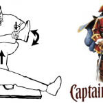
Named after the pose of the trademark pirate for Captain Morgan rum, this technique simplifies the task of pulling the hip back into position. One of the disadvantages of the standard technique is that it takes a fair amount of strength (and patient sedation) to reduce the hip. If the physician is small or the patient is big, the technique may fail.

In the Captain Morgan technique, the patient is left in their usual supine position and the pelvis is fixed to the table using a strap (call your OR to find one). The dislocated hip and the knee are both flexed to 90 degrees. The physician places their foot on the table with their knee behind the patient’s knee. Gentle downward force is placed on the patient’s ankle to keep the knee in flexion, and the physician then pushes down with their own foot, raising their calf. Gentle rotation of the patient’s hip while applying this upward traction behind the patient’s knee usually results in reduction.

Some orthopedic surgeons use a similar technique, but apply downward force on the patient’s ankle, using the leverage across their own knee to develop the reduction force needed. The Captain Morgan technique use the upward lift from their own leg to develop the reduction force. This may be gentler on the patient’s knee.

The authors report a series of 13 reductions, and all but one were successful. The failure occurred due to an intra-articular fragment, and that hip had to be reduced in the operating room. 

I’m interested in hearing comments from anyone who has used this technique (or the leverage one). And does anyone have any other techniques that have worked for them?

Related post:

Reference: The Captain Morgan technique for the reduction of the dislocated hip. Ann Emerg Med (in press) dol:1016/j.annemergmed.2011.07.010, 2011.

Thanks to Sam Stellpflug MD at Regions Hospital for bringing this article to my attention.

Pediatric CT Scans Before Transfer to a Pediatric Trauma Center

CT scan is essential in diagnosing injury, although concerns for unnecessary radiation exposure are growing. These concerns are even greater in children, who may be more likely to have long-term effects from it. This makes avoiding duplication of CT scanning extremely important.

Unfortunately, there are only about 50 pediatric trauma centers in the US, so the majority of seriously injured children are seen at another hospital before transfer. Does CT evaluation at the first hospital increase the likelihood that a repeat scan will be needed at the trauma center, increasing radiation exposure and risk?

Rainbow Babies and Children’s Hospital in Cincinnati looked at 3 years of transfers of injured children from community hospitals. They then looked at how many of those children had an initial head and/or abdomen scan at the outside hospital, and whether a repeat scan of those areas was performed within 4 hours or arrival at Rainbow.

Numbers were small, but here are the results:

  • 33 had an outside CT scan, 28 (90%) were repeated
  • 6 had an outside abdominal scan, 2 (33%) were repeated
  • 55 did not have outside scans, none were repeated at Rainbow. (This is a weird thing to look at. I would hope that the trauma center didn’t have to repeat any of their own scans within 4 hours!)

Bottom line: It is critically important for referring hospitals to use radiation wisely! First, if the patient has obvious injuries that require transfer, don’t scan, just send. If you need to scan to decide whether you can keep the patient, use the best ALARA* technique you can. And trauma centers, please send a copy of your CT protocols to your referring hospitals so they can get the best images possible.

*ALARA = As low as reasonably achievable (applied to radiation exposure). Also known as ALARP outside of North America (as low as reasonably practicable). Click here for more info.

Related posts:

Reference: Computed tomography before transfer to a level I pediatric trauma center risks duplication with associated radiation exposure. J Pediatric Surg 43(12): 2268-2272, 2008.