All posts by The Trauma Pro

What Is: Venous Sinus Thrombosis?

Venous sinus thrombosis is in the news now, since Hillary Clinton is currently hospitalized and being treated for it. Most trauma professionals have probably never heard of it, let alone seen one. Here’s a quick primer on what you need to know about this problem.

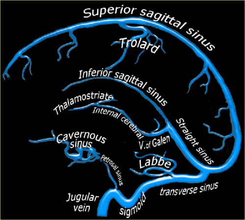
Blood circulation from the cerebral veins drains into the various venous sinus systems around the brain. Additionally, CSF is reabsorbed by the arachnoid villi, which in turn drain into the superior sagittal sinus. A clot in any part of this sinus system (see image below) results in a partial or complete obstruction, which can lead to rapidly increasing pressure in the brain.

image

The condition is very rare, with only about 3 cases per million people recognized. Although most occur in people with clotting abnormalities, trauma is always on the list. The most common traumatic issue is head injury, and almost always involves a skull fracture near the involved venous sinus.

Clinically, awake patients complain of a steadily increasing headache that is not touched by any of the usual analgesics. Some describe a headache with very sudden onset, the so-called “thunderclap headache.” Any number of neurologic abnormalities can then occur, ranging from nothing to seizures to rapid onset of coma.

Diagnosis is difficult. CT scan unreliably shows the lesion, and subtle venous changes may be overshadowed by findings from the original trauma. However, it should be performed first to accurately diagnose the more common problems that are most likely to be present. If the CT comes up clean, the gold standard is MRI with magnetic resonance venography (MRV)

Patients with significant swelling problems may require hyperosmotic therapy and/or decompressive craniectomy. Milder cases can be treated with anticoagulation, but a thorough risk/benefit assessment must be carried out. Remember, these people usually have brain injury that has resulted in bleeding somewhere it shouldn’t be, so anticoagulation can be dangerous! A few rare case reports of thrombolysis (!!) and clot extraction are out there, too, but these are for extreme cases only.

Bottom line: It’s likely that Ms. Clinton suffered a temporal bone fracture from her fall, and did well initially like most patients. However, increasing symptoms (headache), likely prompted a repeat CT followed by the MRI which showed thrombosis of her transverse sinus. Luckily, she did not have significantly increased intracranial pressure leading to more serious neurologic problems. Judicious anticoagulation with warfarin and very close monitoring were instituted, and seem to be working well so far.

References:

  • Thrombosis of the cerebral veins and sinuses. NEJM 352:1791-1798, 2005.
  • Cerebral venous sinus thrombosis complicating traumatic head injury. J Clin Neurosci 19(7):1058-1059, 2012.

Don’t Ignore The Naughty Bits

A major part of any patient encounter is the physical exam. This is particularly true in the trauma patient, because it allows trauma professionals to identify potential life and limb threatening injuries quickly and deal with them. Unfortunately, we tend to mentally block out certain parts of the body, typically the genitalia and perineum, and may not do a complete exam of the area. I call these areas the naughty bits. For those of you who don’t get the reference, here’s the origin of this phrase:

Specifically, the naughty bits are the penis, vagina, perineum, anus and natal cleft (aka the butt crack or arse crack). These areas are more likely to remain covered when the patient arrives, and are less likely to be examined thoroughly.

In penetrating trauma, a detailed exam of these areas is extremely important in every patient to avoid hidden injuries and to determine if nearby internal structures (rectum, urethra) might have been injured.

Here are some tips for each of the areas:

  • Penis – Always look for any blood at the meatus (or a little blood in the underwear) as a possible sign of urethral injury. This is frequently associated with pelvic fractures.
  • Scrotum – Blood staining here is usually from blood dissecting away from pelvic fractures. Patients with this finding are more likely to need angiographic embolization of pelvic bleeding.
  • Vagina – external exam should always be done. Internal and/or speculum exam should be done in pregnant patients, and those with external bleeding or pelvic fractures
  • Perineum – Also associated with pelvic fracture and significant bleeding. Skin tears in this area are usually lacerations indicating an open pelvic fracture. Alert your orthopaedic surgeons early, and do a good, clean rectal exam (carefully wipe away all external blood). Rectal injuries are common with this finding, and a formal proctoscopic will probably be required.
  • Anus – Skin tears virtually guarantee that a deeper rectal injury will be found. Proctoscopic exam in the OR is mandatory.
  • Natal cleft – Usually not a lot going on in this area, except in penetrating injury. This is the only area of the naughty bits that can be safely examined in the lateral position. 

Bottom line: The naughty bits are important because the occasional missed injury in this area can be catastrophic! Do your job and force yourself to overcome any reluctance to examine them.

Related posts:

LifeBot The Next Generation

Over a year ago, I wrote about a product called LifeBot. This technology provides a way to join the ED and prehospital teams as they work on patients. This involves special monitoring equipment in the ambulance (cameras and other telemedicine equipment), a special tablet computing system for data input and imaging, and equipment at the ED base station.

Using the original LifeBot system, medics could relay vitals and EKG data to the base station in real time, receive orders from emergency physicians, and send video feeds and photos from the ambulance.

LifeBot Technology has now released LifeBot 5, the next generation of this system. The unit is now portable, and can be taken out of the ambulance at the scene. It is ruggedized and weighs only 15 pounds, which isn’t bad for field medical equipment. The system now includes a web interface that can mesh with some electronic medical record systems. 

Expect to see more improvements (a defibrillator is slated as the next addition) as well as competing products soon.

What does it cost, you ask? A lot! As always, it’s tough to get exact numbers. The LifeBot 5 should be about $20,000. However, this does not include equipment cost for the base station, which is at least that much, if not more!

Bottom line: Expect further progress in blending the prehospital and emergency department environments. More products like this will become available, extending the senses of emergency physicians and providing additional assistance to prehospital providers.

Related post: The “super ambulance” of the future

Website: http://www.lifebot.us/dreams/

Disclosure: I have no financial interest in Lifebot Technology

A Cool Way To Look At Injury Data

Governmental agencies everywhere collect trauma related data. The US federal government maintains a number of databases, such as the Fatal Accident Reporting System (FARS), the Census of Fatal Occupational Injuries (CFOI) and many others. States collect similar but smaller datasets. Even towns and municipalities collate injury information in the form of prehospital run sheets.

But reams of data are of no use unless you can learn something from it. Unfortunately, most of this data is tucked away in database management systems, or in some cases just stacks of paper forms locked up somewhere. In order for humans to make sense of it and do useful things with it, we need to transform it into forms that we can easily interpret and make sense of. 

Fortunately, there are lots of visual, electronic tools available to help us do just that. One of the most helpful tools is the programmable geographic information system (GIS). An example of this is Google Maps. Most of us have used this or a similar tool in some form, usually to get directions from here to there. But you may not be aware that Google provides a programming interface so a savvy user can place any type of geography-related data on the map, creating what is called a mashup.

Imagine crossing the FARS database, which contains extensive data points on every fatal road accident in the US, with a mapping system. This would allow creation of a map showing where every person lost their life in a road accident, along with additional pertinent information about the event. A great example of this is demonstrated below. It was created by ITO World Ltd., based in the UK. They crossed fatality information with geographic map data in both the US and the UK.

This map shows fatal road events around Minneapolis from 2001 to 2009. The type of event (pedestrian struck, motor vehicle crash, etc.) is displayed along with age, year and sex. It is movable and zoomable so it can be viewed it in great detail. Click on the map above to open a new window to the full map.

Bottom line: Using trauma data / map mashups is a great way to visualize complex information. It also allows us to plan meaningful prevention activities based on local information (a requirement for ACS trauma center verification). Imagine looking over such a map of your city, and identifying a cluster of pedestrian fatalities. Then you notice that this cluster is 2 blocks away from an elementary school. This could prompt you to work with the school to implement automobile awareness programs for the children, have the city review signage and obstructions to view in the area, and optimize the number and placement of crossing guards. Then redo the map afterwards to judge the impact. Wow!

Website: http://map.itoworld.com/road-casualties-usa#fullscreen 

Reference: Using geographic information systems in injury research. J Nurs Scholarsh 39(4):306-311, 2007.