All posts by The Trauma Pro

To Probe or Not To Probe: Penetrating Wounds

There is considerable variability in the way that penetrating wounds are approached. Some are located over areas of lesser importance (distal extremities) or are so superficial that they obviously don’t fully penetrate the skin.

Unfortunately, some involve high-value structures (much of the neck and torso), or are too small to tell if they penetrate (ice pick injury). How should these injuries be approached?

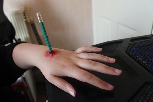
Too often, someone just probes the wound and makes a pronouncement based on that assessment. Unfortunately, there are major problems with this technique:

  • The tract may be too small to appreciate with a finger or even a cotton-tip swab
  • The tract may be oriented in an unexpected direction, or the soft tissues may have moved after the penetration occurred. In this case, the examiner may not appreciate any significant depth to the wound.
  • Inserting an object may violate a structure that you wish it hadn’t (resulting in a hissing sound after probing a chest wound, or a column of blood after probing the neck)

A better way to approach these wounds is as follows:

  • Is the patient unstable? If so, you know the penetration caused the problem and the patient belongs in the OR.
  • Is there other evidence of deep injury, such as peritonitis with a penetrating abdominal wound? If so, the patient still needs to go to the OR.
  • Do a legitimate local wound exploration. This entails making the hole bigger with a knife, and using surgical instruments and your eyes to find the bottom of the tract. Obviously, there are some parts of the body where this cannot be done, such as the face, but they probably don’t need this kind of workup anyway.

As one of my mentors, John Weigelt, used to say, “Doctor, do you have an eye on the end of your finger?” In general, don’t use anything that doesn’t involve an eyeball in your local wound explorations!

Taking X-Rays Through A Backboard

Most major trauma patients are transported to the trauma center on a backboard. And nearly all of them get at least a chest x-ray, and possibly a pelvis x-ray. But do the backboard and x-rays mix?

There seems to be a debate in our ED about the quality of x-rays obtained through a backboard. This has led to a push to hold the x-ray until the patient has been rolled and the board removed. Unfortunately, since we must wait and actually view the image before transporting the patient, this can lead to a delay in leaving the resuscitation room.

So what’s the problem? Most backboards are made of plastic these days, with very few metal parts. Even the strap buckles are usually plastic. And plastic is, for the most part, transparent to x-rays. Yes, if the board is thick it may lighten the image a bit. And if there are cutouts in it, they may show as darker areas. However, they tend to have smooth and regular borders that are easily distinguished from structures in the human body.

Bottom line: The densities and irregularities in backboards do not significantly degrade x-ray images. Remember, you are looking for large collections of air (pneumothorax) or fluid (hemothorax) in the chest, and major bony disruption in the pelvis. These are easily seen through a backboard, or even if parts of the body are off the edge of the image. Don’t wait to get those x-rays, have them done as early as possible so you can view them and move on to the next phase of care.

Related posts:

The Prehospital “Nonstandard Patient Position” Sign

Prehospital providers follow protocols for securing and transporting trauma patients. These may include cervical spine stabilization and short or long backboards. Every once in a while they can’t follow protocol, and in my experience it usually means that something is very wrong. 

There are three typical problems leading to nonstandard transport positions:

  • Occult airway injury – These patients have either blunt injury to the neck, smoke inhalation, or penetrating injury to the submandibular area. They tend to have problems protecting their own airway when they are supine, so they insist on being transported in an upright position.
  • Impalement – Since the general rule is to leave foreign objects in place to avoid potential bleeding, the patient is positioned in an odd way to accommodate both them and the impaling object. 
  • Life-threatening bleeding – Patients with exsanguinating hemorrhage who are awake tend to insist on transport in certain positions. Most with serious chest hemorrhage complain that they can’t breathe and want to sit upright. Those with severe pelvic fractures complain of pelvic or back pain and may prefer lying on their side during transport.

Bottom line: If prehospital providers bring a trauma patient to you in a non-supine position, be very afraid. If not done already, activate your trauma team. Talk to the medics to find out why they had to use a nonstandard position. Then rapidly assess the patient to rule out life-threatening issues.


Related posts:

Don’t Get Lateral View Chest Xrays to Diagnose Pneumothorax

Pneumothorax is typically diagnosed radiographically. Significant pneumothoraces show up on chest xray, and even small ones can be demonstrated with CT.

Typically, a known pneumothorax is followed with serial chest xrays. If patient condition permits, these should be performed using the classic technique (upright, PA, tube 72" away). Unfortunately, physicians are used to ordering the chest xray as a bundle of both the PA and lateral views. 

The lateral chest xray adds absolutely no useful information. The shoulder structures are in the way, and they obstruct a clear view of the lung apices, which is where the money is for detecting a simple pneumothorax. The xray below is of a patient with a small apical pneumothorax. There is no evidence of it on this lateral view.

Bottom line: only order PA views (or AP views in patients who can’t stand up) to follow simple pneumothoraces. Don’t fall into the trap of automatically ordering the lateral view as well!

Lateral chest xray

Crowdsourcing Medical Research

Medical resource is hard to do. It’s tough to come up with an idea no one has explored, design the study, accumulate subjects (for clinical research), analyze it, and then write a good paper. You’re probably familiar with my lamentations over all of the small,retrospective studies that seem to dominate the medical literature.

Crowdsourcing takes advantage of the “human computing power” of ordinary people all around us. Some of you may remember the protein folding computer game that was distributed for free about 10 years ago, allowing everyone to try their hand at designing protein configurations. Turns out, masses of regular people are better than computers for doing this kind of stuff. And the results were impressive. Or look at the power of Amazon’s Mechanical Turk, a crowdsourcing platform for a variety of tasks. Or KickStarter for funding projects that would have a tough time getting money on their own.

Researchers at Penn used crowdsourcing in a study to map the locations of all automated external defibrillators in Philadelphia. They called it the MyHeartMap challenge. The crowd quickly identified and catalogued over 1400 of them. They are now using crowdsourcing to perform literature sources to collect and analyze health-related studies using free medical literature search sites.

The crowd does best on studies involving problem solving, data processing, monitoring and surveying. The downside is that there is built-in variability when using the crowd, which can make it difficult to replicate and confirm validity. But the sheer numbers that can be accumulated are far larger than what can be expected using traditional research methods.

Bottom line: Crowdsourced research has significant potential to accelerate and improve the quality of medical research (and in other fields as well). Anyone engaged in research needs to look at their own projects to determine if any of them can be improved using crowdsourcing. Look at what crowds are good at (solving problems, observing and reporting), and use them to bolster and improve our knowledge base.

Reference: Crowdsourcing – Harnessing the masses to advance health and medicine, a systematic review. J General Int Med DOI: 10.1007/s11606-013-2536-8, 2013.