All posts by The Trauma Pro

Clinical Manifestations Of Fat Embolism Syndrome

There are three organ systems that are classically involved in FES: pulmonary, CNS, and skin. Manifestations generally begin between 24 and 72 hours after injury. In rare cases, symptoms can begin within 12 hours. In my experience, these tend to be the ones that become the most severe and are frequently life-threatening.

Pulmonary (95% of cases): This is the most common manifestation of FES, and may occur without other signs and symptoms. Nearly all patients develop some degree of hypoxia. Progressive tachypnea and mild tachycardia may provide the first clinical clue if oxygen saturation is not being monitored.

Chest x-ray is usually unremarkable early on. And once the syndrome has developed, it is generally not helpful. CT scan is useful for defining the extent of pulmonary injury, but lags the clinical picture by several days. Findings are non-specific, usually consisting of small, ground-glass opacities in the periphery.
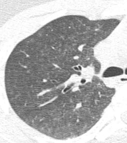
In the example above, the opacities are very small and difficult to see.
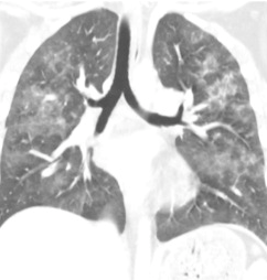
But they’re a little more obvious here!

Other CT findings include small pulmonary nodules in the upper lobes or along peripheral pulmonary vessels. These are thought to be areas of obstruction caused by the emboli. Nonspecific pleural effusions may be seen, and bronchial thickening has also been described. Rarely, fat globules may be seen in the lower extremity veins or IVC, and should immediately raise suspicion for developing FES even before symptoms develop.

CNS (60% of cases): If they occur, CNS changes generally crop up after the pulmonary manifestations begin. Generally, they start as mild confusion, but can progress to decreasing level of consciousness and even coma. Focal neurologic deficits are occasionally seen, and seizures can occur.

The actual mechanism behind this appears to be very similar to the skin changes which will be described in the next section. Emboli occur in vessels predominantly in the white matter of the brain. This leads to petechial hemorrhages, which are likely due to the inflammatory mechanisms previously described.
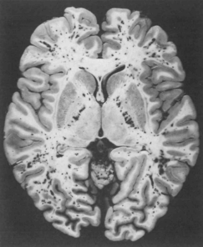
Note the numerous dark petechiae visible in the white matter in this specimen.

Retinal exam can also show evidence of fat embolism. Fat globules may actually be seen in the retinal vessels early.
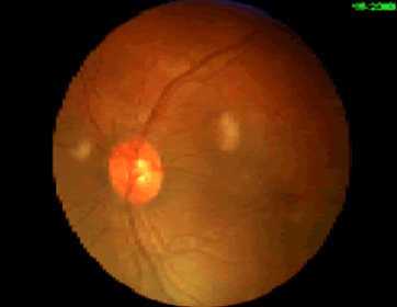
Note the fat globules at the 9:30 and 2:00 positions to the optic nerve in the image above.

Skin (33% of cases): The most recognizable sign of FES is the petechial skin rash. This rash usually involves the torso, and axillary petechiae are very common. It can spread to involve the head and neck, and occasionally the extremities. Subconjunctival hemorrhages are sometimes seen. The rash tends to be transient and usually lasts only a few days. Here is an example of the classic petechial rash.

Other findings: Fat globules may be found in the urine in patients with FES. However, they are commonly present in patients with long bone fractures, so their presence is not helpful or predictive. Nonspecific findings such as fever, leukocytosis, anemia, and thrombocytosis are also relatively common. In severe cases, cardiac dysfunction, hypotension, and peripheral hypoperfusion can occur. I have personally seen necrosis of fingers and toes from a very severe case.

Unfortunately, the “classic” triad of mental status changes, skin rash, and pulmonary insufficiency are seen in only a small minority of patients. Typically, only one or two signs and symptoms appear at the same time, making diagnosis a bit challenging.

In the next post, making the diagnosis of fat embolism syndrome.

Fat Embolism vs Fat Embolism Syndrome

It’s fat embolism week! I’ll cover this uncommon, yet very important clinical condition in my next four posts.

Fat embolism syndrome (FES) is one of those clinical problems that trauma professionals read about during their training, then rarely ever see. Although the clinical manifestations are frequently mild, they can progress rapidly and become life-threatening. Over the next five days, I’ll try to  help you better understand this condition, and provide details on diagnosis and treatment.

Fat embolism syndrome (FES) is a constellation of findings that arise from a single, unified cause: the escape of fat globules into the circulation (fat embolism). The ultimate resting places of those globules determine the specific manifestations of FES seen in clinical practice. When it occurs, it typically becomes apparent 24 to 72 hours after injury.

Simple fat embolism occurs to some degree any time tissues containing fat are manipulated or injured. It has been demonstrated during plastic surgical injections for cosmetic purposes and lipid infusions. It is more frequently seen with orthopedic injuries, especially those involving the femurs and pelvis. And it makes sense that the more fractures that are present, the more likely fat embolism will occur. Embolism is also known to occur when performing orthopedic procedures, particularly those involving the marrow cavity (intramedullary nailing), but has also been reported in total knee and hip procedures.

Fat embolism syndrome has a generally reported incidence of 1 – 10%, although I believe that is on the high side. I see a case every 3 – 4 years in a predominantly blunt, fracture-laden practice. Fat embolism without symptoms occurs much more frequently. A study from 1995 using transesophageal echo found evidence of emboli in 90% of patients with long bone fractures.

But how do these fat globules get into the circulation and produce such chaos? We know that they can be mechanically pushed into small venules when tissues containing fat cells or bone marrow are injured. In bone, there are numerous small venules located throughout that are anchored to it. When the bone is fractured, these venules tear and are held open so yellow (fatty) marrow can be pushed into them.

If enough emboli enter the blood stream, they may accumulate in the end vessels of tissues and block flow. Although this is a simple and appealing explanation, it may not be the full story. If the emboli primarily occur during and after injury, why does it take several days for the full-blown syndrome to develop?

A likely explanation is that the fat globules begin to degrade while in the circulatory system. Breakdown into free fatty acids results in the release of a cascade of cytokines and other mediators. The inflammatory response around the end vessels create the gross pathology that we associate with fat embolism syndrome.

In the next post, clinical manifestations of fat embolism syndrome.

Are Transfusing Too Much Blood During The MTP?

The activation of the massive transfusion protocol (MTP) for hypotension is commonplace. The MTP provides rapid access to large volumes of blood products with a simple order. Trauma centers each design their own protocol, which usually includes four to six units of PRBC per MTP “pack.”

This rapid delivery system, coupled with rapid infusion systems, allows the delivery of large volumes of blood and other blood products very quickly. But could it be that this system is too slick, and we are a bit too zealous, and could even possibly transfuse too much blood?

The trauma group at Cedars-Sinai in Los Angeles retrospectively reviewed their own experience via registry data with their MTP over a 2.5 year period for evidence of overtransfusion. All patients who received blood via the MTP were included. Patients who had a continuous MTP > 24 hours long, those who died within 24 hours, and those who had a missing post-resuscitation hemoglobin (Hgb) were excluded.

The authors arbitrarily defined overtransfusion as a Hgb > 11 at 24 hours. They also compared the Hgb at the end of the MTP and upon discharge with this threshold. They chose this Hgb value because it allows for some clinical uncertainty in interpreting the various endpoints to resuscitation.

Here are the factoids:

  • 240 patients underwent MTP during the study period, but 100 were excluded using the criteria above, leaving 140 study patients
  • Average injury severity was high (24) and 38% suffered penetrating injury
  • Median admission Hgb was 12.6
  • At the conclusion of the MTP, 71% were overtransfused using the study definition, 44% met criteria 24 hours after admission, and 30% did at time of discharge
  • Overtransfused patients were more likely to have a penetrating mechanism, lower initial base excess, and lower ISS (median 19)

The authors concluded that overtransfusion is more common than we think. This may lead to overutilization of blood products, which has become much more problematic during the COVID epidemic. They recommend that trauma centers track this metric and consider it as a quality of care measurement.

Bottom line: This is a nicely crafted and well-written study. It asks a simple question and answers it with a clear design and analysis. The authors critique their own work, offering a comprehensive list of limitations and a solid rationale for their assumptions and conclusions. They also offer a good explanation for their choice of Hgb threshold in defining overtransfusion.

I agree that overtranfusion truly does occur, and I have seen it many times first-hand. The most common reason is the lack of well-defined and reliable resuscitation endpoints. How do we know when to stop? What should we use? Blood pressure? Base excess? TEG or ROTEM values? There are many other possibilities, but none seem reliable enough to use in every patient. 

Patients with penetrating injury proceeding quickly to OR more commonly experience overtransfusion. This may be due to the reflexive administration of everything in each cooler and the sheer speed with which our rapid infuser technology can deliver products. The more product in the cooler, the more that is given, which may lead to the overtranfused condition. 

The authors suggest reviewing the makeup of the individual MTP packs, and this makes sense. Are there too many in it? This could be a contributing factor to overtransfusion. It might be an interesting exercise to do a quick registry review at your own center to obtain a count of the number of MTP patients with a final Hgb > 11. If you find that your numbers are high, consider reducing the number of red cell packs in the cooler to just four. But if you already only include four, don’t reduce it any further. And in any case, critically review the clinical indicators your  surgeons use to decide to end the MTP to see if, as a group, they can settle on one to use consistently. 

Reference: Overtransfusion of packed red blood cells during massive transfusion activation: a potential quality metric for trauma resuscitation. Trauma Surg Acute Care Open 7:e000896., July 26 2022.

Trauma Patient Transport By Police, Not EMS

When I was at Penn 30+ years ago, I was fascinated to see that police officers were allowed to transport penetrating trauma patients to the hospital. They had no medical training and no specific equipment. They basically tossed the patient into the back seat, drove as fast as possible to a trauma center, and dropped them off. Then they (hopefully) hosed down the inside of the squad car.

Granted, it was fast. But did it benefit the patient? The trauma group at Penn decided to look at this to see if there was some benefit (survival) to this practice. They retrospectively looked at 5 years of data in the mid-2000’s, thus comparing the results of police transport with reasonably state of the art EMS transport.

They found over 2100 penetrating injury transports during this time frame (!), and roughly a quarter of those (27%) were transported by police. About 71% were gunshots vs 29% stabs.

Here are the factoids:

  • The police transported more badly injured patients (ISS=14) than EMS (ISS=10)
  • About 21% of police transports died, compared to 15% for EMS
  • But when mortality was corrected for the higher ISS transported by police, it was equivalent for the two modes of transport

Although they did not show a survival benefit to this practice, there was certainly no harm done. And in busy urban environments, such a policy could offload some of the workload from busy EMS services.

Bottom line: Certainly this is not a perfect paper. But it does add more fuel to the “stay and play” vs “scoop and run” debate. It seems to lend credence to the concept that, in the field, less is better in penetrating trauma. What really saves these patients is definitive control of bleeding, which neither police nor paramedics can provide. Therefore, whoever gets the patient to the trauma center in the least time wins. And so does the patient.

Related posts:

Reference: Injury-adjusted mortality of patients transported by police following penetrating trauma. Acad Emerg Med 18(1):32-37, 2011.

Fracture Care Of The Future: Traditional Casts vs 3D-Printed Braces

I’ve been fascinated by 3D printing for at least a decade.  Here are some examples from previous posts:

Unfortunately, practical applications have been relatively limited in the field of trauma.  But a lot has been going on in the background. The trauma research group at Erasmus Medical Center in Rotterdam recently published a systematic review on very practical work using 3D printing to produce casts and splints.

Sounds like a very mundane problem to through high tech at, right? But for those of you who look after patients with fractures that have been casted, you know the problems that can arise. Casts can be too tight. They can be ill-fitting. The patient may have soft tissue injuries that require windows cut into the side of the cast. Additional technology such as electrical stimulators may be indicated to enhance healing.

The old-fashioned way of creating a plaster or fiberglass cast seems crude. It is shaped by hand using skill and a fair amount of guesswork. If it’s just a bit too tight, serious complications may occur. If windows are not cut properly, it can destabilize the entire cast.

The Rotterdam trauma research group performed a systematic review of 12 papers that have been published on the topic of 3D-printed casts used in the treatment of forearm fractures. The authors found that most currently use a technique called fused deposition modeling with a polylactic acid substrate.

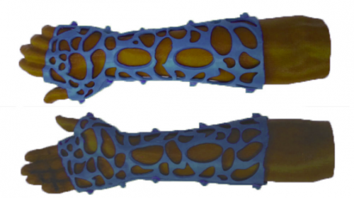
Instead of relying on subjective skill and luck to shape the brace, the uninjured forearm is scanned with a 3D scanner. The data is fed to a computer aided design (CAD) workstation and a mirror image is created and further refined. Special features such as soft tissue windows or entry points for bone stimulators can be designed into the brace at that time. Because the strength of polycarbonate exceeds that of plaster and fiberglass, it is possible to create a design with a great deal of open area so the underlying skin can be monitored. And allowances can be made for areas with swelling not present on the control extremity.

The data is then fed to a 3D printer to actually create the cast. Here’s an example:

This design is stronger that a traditional cast, is cool and comfortable, and avoids problems with hidden tissue injury or unrecognized foreign objects dropping into the cast creating major problems.

The use of 3D-printed casts and braces is relatively new and is used in only a few centers. For this reason, we do not have enough numbers to show that it is equivalent to traditional casting. Yet. But as the price continues to drop and use becomes more widespread, it’s only a matter of time before you start seeing these items in your own trauma center.

Reference: Personalize d 3D-printed forearm braces as an alternative for a traditional plaster cast or splint; A systematic review. Injury, in press, July 29, 2022. https://doi.org/10.1016/j.injury.2022.07.020