All posts by The Trauma Pro

Prevention: Handlebar Injury

“Necessity is the mother of invention”

                                   -Unknown

I’ve managed several cases of injury due to bicycle handlebars over the years. Typically, a smaller child crashes his or her bike, and the handlebar hits them in the epigastrium. Children have thinner abdominal walls and less developed muscular to protect them, so this very focal impact can do a lot of damage. 

There is now a clever and inexpensive solution available that can decrease the number of injuries we see from this common mechanism. It’s called the Handlebar Helmet, and was developed by the parents of a 4 year old boy who suffered this injury. It is essentially a special plastic cap that fits on the end of the handlebars. It’s designed to diffuse the pressure of any impact with the handlebar. This product actually does double duty, protecting during a crash, and also preventing injury if a child trips and falls on a bike that is lying down.

image

image

The product is very easy to install, and comes in multiple colors so it can be “cool” (very important to kids). This is a nice, simple idea that can prevent potentially devastating injuries.

Over the next two days, I’m going to rerun an interesting pediatric case of handlebar injury.

Reference: The Handlebar Helmet. www.handlebarhelmet.com

Note: I have no financial interest in this product.

Where Do Pulmonary Emboli Really Come From?

For a long time, we “knew” that pulmonary emboli were a possible and dreaded complication of deep venous thrombosis (DVT). However, we are beginning to discover that this is not always the case. The group in San Diego decided to see if there really are two different types of PE in trauma, and what that means.

Scripps Mercy Hospital, a level I trauma center, looked at 5 ½ years of their experience with adult trauma patients who were routinely screened for DVT. Any of these patients who developed a PE within 6 weeks of admission were evaluated further.

Here are the factoids:

  • Duplex screening from groin to ankle was carried out twice weekly in ICU patients, and once weekly in ward patients
  • Surveillance was carried out if the patient would be non-ambulatory for more than 72 hours, or were at moderate or higher risk for DVT using the ACCP guidelines
  • Nearly 12,000 patients were evaluated by the trauma service and 2,881 underwent surveillance
  • 31 patients (1%) developed a PE
  • 12 of these 31 had DVT identified before or immediately after their PE. Clot was below-knee in 9 (!), above-knee in 2, and in the IJ in one.
  • 19 patients had PE but no DVT identified (de novo PE, DNPE)
  • DNPE tended to be single and peripherally located, and associated with rib fractures, pulmonary contusions, blood transfusions, and pneumonia
  • DVT + PE were more often found in multiple lobes or bilaterally

Bottom line: Like most, this is not a perfect study, but it’s a really good one. It is looking more and more likely that some PEs arise de novo, without any associated DVT. These clots are more likely to be linked to some type of inflammatory process, and have a tendency toward causing more of the classic signs and symptoms of PE. There are still lots of questions to be answered, like do you need to anticoagulate the de novo PEs? But for now, no change in practice. Just be aware that these might not be as bad as they seem.

Related posts:

Reference: Pulmonary embolism without deep venous thrombosis: de novo or missed deep venous thrombosis? J Trauma 76(5):1270-1281, 2014.

When Is It Too Late To Call A Trauma Activation?

Admit it. It’s happened to you. You get paged to a trauma activation, hustle on down to the ED, and get dressed. The patient is calmly and comfortably lying on a cart, staring at you like you’re from Mars. Then you hear the story. He has a grade V spleen injury. But he just got back from CT scan. And his car crash was yesterday

Is this appropriate? The answer is no! But, as you will see, the answer is not always as obvious as this example. The top thing to keep in mind in triggering a trauma activation appropriately is the reason behind having them in the first place.

The entire purpose of a trauma activation is speed. The assumption must be that your patient is dying and you have to (quickly) prove that they are not. It’s the null hypothesis of trauma.

Trauma teams are designed with certain common features:

  • A group of people with a common purpose
  • The ability to speed through the exam and bedside procedures via division of labor
  • Rapid access to diagnostic studies, like CT scan
  • Availability of blood products, if needed
  • Immediate access to an OR, if needed
  • Recognition in key departments throughout the hospital that a patient may need resources quickly

Every trauma center has trauma activation triage criteria that try to predict which patients will need this kind of speed. Does the patient in the example above need this? NO! He’s already been selected out to do well. Why, he’s practically finished the nonoperative solid organ management protocol on his own at home.

Here are some general rules:

  • If the patient meets any of your physiologic and/or anatomic criteria, they are or can be sick. Trigger immediately, regardless of how much time has passed.
  • If they meet only mechanism criteria and it’s been more than 6 hours since the event, they probably do not need the fast track.
  • If they only meet the “clinician / EMS judgment” criteria, think about what the suspected injuries are based on a quick history and brief exam. Once again, if more than 6 hours have passed and there are no physiologic disturbances, the time for needing a trauma activation is probably past.

If you do decide not to trigger an activation in one of these cases, please let your trauma administrative team (trauma medical director, trauma program manager) know as soon as possible. This may appear to be undertriage as they analyze the admission, and it’s important for them to know the reasoning behind your choice so they can accurately document under- and over-triage.

Related posts:

What To Do? Small Hospital, Unstable Patient

It’s the situation that physicians in smaller hospitals dread. A major trauma patient gets dropped off at the door. You do your evaluation, quickly determining that they need services that you don’t just have (head injury and positive FAST in the abdomen, let’s say). You call your community EMS service to transport to a Level I trauma center, which is about 30 minutes away by ground. And just as they are rolling out the door to the rig, the blood pressure drops to 60! What to do?!

The ATLS course is very clear, and very correct. Back into the ED for a quick re-evaluation. The most common cause for a significant disturbance in vitals or exam lies within the primary survey. You will almost always find a problem with Airway, Breathing, or Circulation. (A Disability problem can cause a problem on rare occasion (hypotension from impending herniation), but there’s not much that you can do about it, really. Hyperventilate, hyperosmolar therapy, okay but probably a poor outcome for the patient anyway.)

So you didn’t find any airway or breathing issue. But the abdominal stripe(s) you saw on FAST are larger, so it’s circulation. Now what? And does it matter if you have a surgeon available on call? The answer is simpler than you think.

ATLS says that, if you have surgical support available you have to use it in this type of situation. If you don’t have it, package the patient with a lot of blood and plasma and send. If you have a physician or nurse to spare you could consider sending them along to help during transport, but for small community hospitals this is not practical.

But if you do have a surgeon, does it make sense to use them? Not always! You must take into account response times and transport times. Let’s say it’s 2:00 am and you call your surgeon for this hypotensive patient. They may take up to 30 minutes to get in and see the patient. They then agree that the patient needs a laparotomy and she proceeds to call in the OR team. Yet another 30 minutes tick by.  Will the patient still even be alive when they roll into the OR?

Or you could just put the patient back in the ambulance (air preferably, but ground if you have to) and get them to your trauma center quickly. They can then be whisked directly into a waiting OR in less than 30 minutes from your door. This is probably the ideal solution here. Obviously this doesn’t work as well if you are a few hours away from your resource trauma center. 

Bottom line: Deciding what to do with a patient that needs urgent treatment that you can’t immediately deliver is tough! That’s why it’s always a calculus problem when you’re faced with this situation. But take all of the response and transport times into account, and do what’s best for your patient! 

Nail In The Neck: A Novel Removal Option

Here’s a post from my archive describing a different way to remove the foreign body. This is the technique I used, instead of the standard neck incision. The final incision was just a slight extension of the puncture wound, measuring only 1cm. I was able to grasp the head and pull it out without difficulty. The surprising thing to me was the amount of force I needed to apply to actually pull it out! No bleeding, no problems. The patient was observed for 24 hours and discharged home. He had no complications.

A Cool Way To Remove Embedded Foreign Bodies

Many of us have had the experience of digging into bloody tissue for long periods of time trying to locate the object, even with fluoroscopy. Well, there’s a better way of doing this.

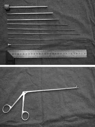
A group in China described a technique using a fancy form of needle localization. They employed a set of instruments normally used for lumbar diskectomy (see photo). This set includes a long 18 Ga needle with a removable hub, several dilators and an outer cannula with a 5.8mm diameter. A pair of 3.8mm grasping forceps is also used.

The foreign body is located using a C-arm fluoroscopy unit and the best approach is planned. The 18 Ga needle is then inserted using fluoro until it touches the object. The hub is removed and dilators are inserted over the needle, one after the other. The outer cannula is then placed over them, and the needle and dilators are then removed. The cannula is manipulated until the foreign body (or a part of it) is located within the cannula. It is then grasped and removed, along with the cannula if needed. If the object is too large to enter the cannula, the cannula is pulled back slightly and the grasper introduced past the end of it to grip and remove the foreign body.

The writers shared the details of 76 patients who had a total of 251 foreign bodies removed over a 6 year period. The depth varied from 2.5 to 8.5cm. Procedure time ranged from 8 to 15 minutes, and fluoro exposure varied from 1 to 4 minutes. Success rate was 100% (all foreign bodies were removed) and there were no complications.

Bottom line: This is a very slick technique that promises to dramatically increase the success rate and decrease complications from removing foreign bodies. The amount of time spent is much less than the brute force technique, as is the amount of soft tissue trauma. Large objects that cannot be grasped with these forceps cannot be removed with this method. Although I am a little concerned that the authors’ results were so perfect, it’s certainly worth a try!

Related posts:

Reference: Percutaneous extraction of deeply-embedded radiopaque foreign bodies using a less-invasive technique under image guidance. J Trauma 72(1):302-305, 2012.