All posts by The Trauma Pro

Acute Ethanol Intoxication And The Banana Bag

It’s one of those time honored treatments that most hospital-based providers are familiar with. The banana bag, reserved for intoxicated patients presenting to the ED or admitted to the hospital. They’ve been around so long, we just take them for granted. But like most things that have become dogmatic, they are due to be questioned from time to time.

image

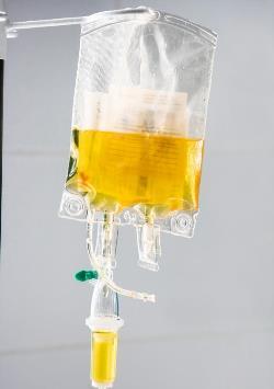
A banana bag is a proprietary mix of “good” stuff, including electrolytes and vitamins, especially thiamine and magnesium. The exact content varies from hospital to hospital. Thiamine and other B vitamins give the resulting solution the characteristic color, hence the term “banana.”

Does it actually do good things like ward off Wernicke’s encephalopathy and megaloblastic anemia? A paper from Jacobi Medical Center in the Bronx prospectively evaluated a series of intoxicated people entering their ED. They drew vitamin B12, folate, and thiamine levels to see if they were deficient enough to even need vitamin supplementation.

Here are the factoids:

  • These folks (only 77 patients) were very drunk! Average BAC was 280mg/dL.
  • Vitamin B12 and folate levels were not critically low in any patient
  • Thiamine was low in 15% of patients, but none had clinical evidence of a deficiency
  • Later review of prior visits revealed that some patients with low levels had received a previous banana bag within 1 month. Did it do any good?

Bottom line: Most of our intoxicated patients are not vitamin deficient, and don’t need supplementation. The real kicker is that we almost never really try to find out if the patient might be a chronic abuser and potentially at risk. We just hang the bag. Remember, everything we do in medicine has a potential downside. And if the patient really doesn’t need a banana bag in the first place, there is no benefit to balance that risk. The next time you ask for that little yellow bag, think again!

Reference: Vitamin deficiencies in acutely intoxicated patients in the ED. Am J Emerg Med 26(7):729-795, 2008.

The Flat Vena Cava Sign? Published and Revisited

I’ve previously blogged about the flat vena cava sign as an indicator of low volume status in trauma patients. And I commented on this paper when it was presented at EAST, which had a surprisingly negative result. It’s now been vetted by peer reviewers and published, and I’ve had the opportunity to read through the entire manuscript (always important). So let’s take a second look now.

A retrospective study at George Washington University was carried out over a one year period. They looked at all of their highest level trauma activation patients who also underwent CT scan of the abdomen. Images were read by three radiologists and inter-rater reliability was reviewed. The transverse to anteroposterior diameter ratios were calculated to determine flatness.

Here are the factoids:

  • 276 patients met enrollment criteria, and were mostly male and blunt trauma
  • The IVC was nearly round in 21% of patients and collapsed in 26%
  • There was no association between IVC shape and shock index, blood pressure, Hbg, lactate, urgent operation, angiography or length of stay
  • There was also no association between IVC shape and blood transfusion or death
  • Correlation of the reads between radiologists was good

So what gives? A paper I reviewed three years ago in the Journal of Trauma came to a different conclusion. They found that a flat IVC on CT scan (defined as a transverse to AP ratio of 4:1 or greater) was associated with a significantly higher chance of receiving more crystalloid or blood, as well as requiring an operation within 24 hours. 

This newer paper was able to look at a larger group of patients, and they were able to tease out why it initially looked like the flat cava looked like a good predictor for bad things to come. The problem was statistical skewing from a few extreme outliers. When properly corrected, it completely changed things. And looking at the older study, it appears that outliers may have also been the reason for the positive result. This is why I encourage everyone to always read the entire paper! The older paper involved a smaller series (114 patients), but it was prospective and seemed to have reasonable statistical analyses.

image

Bottom line: It looks like the flat vena cava sign, as measured by a static CT, should be discarded as an indicator of impending shock. Whether or not a more dynamic look (using ultrasound) is valuable remains to be determined.

Related post:

Reference: Inferior vena cava size is not associated with shock following injury. J Trauma 77(1):34-39, 2014.

Phlebotomy And Pediatric Solid Organ Injury

I recently wrote about this journal article from a couple of pediatric trauma programs in New York. The article tried to focus on reducing the rate of phlebotomy in children who are being observed for solid organ injury. I was more excited about the overall protocol being used to manage liver and spleen injury, as it was a great advance over the original APSA guideline. But let’s look at the phlebotomy part as well.

This is an interestingly weird study, and you’ll see what I mean shortly. Two New York trauma hospitals that take care of pediatric patients pooled 4 years of registry records on children with isolated blunt liver and/or spleen injuries. Then they did a tabletop excercise, looking at “what if” they had applied the APSA guideline, and “what if” they had applied their new, proposed guideline.

Interestingly, this implies that they were using neither! I presume they are trying to justify (and push all their partners) to move to the new protocol from (probably) random, individual choice.

Here are the factoids:

  • 120 records were identified across the 2 hospitals that met criteria
  • Late presentation to the hospital, contrast extravasation, comorbidities, lack of imaging, operative intervention at an outside hospital excluded 59 patients, leaving 61 for analysis. Three of those patients became unstable and were also excluded.
  • None of the remaining patients required operation or angioembolization
  • Use of the “new” (proposed) protocol would reduce ICU admissions by 65%, reduce blood draws by 70%, and reduce hospital stay by 37%
  • Conclusion: use of the protocol would eliminate the need for serial phlebotomy (huh?)

Bottom line: Huh? All this to justify decreasing blood draws? I know, kids hate needles, but the data on decreased length of stay in the hospital and ICU is much more important! We’ve been using a protocol similar to their “new” one at Regions Hospital, which I’ve shared below. We’ve been enjoying decreased resource utilization, blood draws, and very short lengths of stay for over a decade. And our analysis showed that we save $1000 for every patient entering the protocol, compared to the old-fashioned and inefficient way we used to manage them.

Related posts:

Reference: Reducing scheduled phlebotomy in stable pediatric patients with liver or spleen injury. J Ped Surg 49(5):759-762, 2014.

Pediatric Solid Organ Injury Management: It’s About Time!

There was an interesting article released in the Journal of Pediatric Surgery in May about spleen and liver injury management in children. It’s interesting because if you just look at the title, you might just skip over it. The title suggests that it describes reducing scheduled phlebotomy in kids who are undergoing solid organ injury management. But the real meat of this article has to do with the protocol they are using to treat the children.

Nonoperative management of these injuries in children started becoming popular 40 years ago (!). But for decades, everyone put their own spin on how to do it. Bed rest for a week (or more). NPO for days! Limited physical activity for extended periods. Then the American Pediatric Surgery Association (APSA) published a set of guidelines about 15 years ago that took some of the guesswork out of it.

Although nonoperative management of these injuries in kids preceded its adoption in adults by a nearly two decades, it has languished in the APSA format for quite some time. Many pediatric surgeons still use these guidelines, even though adult spleen and liver injury management have advanced to shorter and more streamlined care.

We adopted a solid organ management guideline at Regions Hospital over ten years ago, and have made a few minor tweaks over the years. Nowadays, our grade I-III injuries can be home as early as 36 hours after admission, and frequently are. Grades IV and V are eligible to be discharged after just 24 more hours if they have no other injuries to keep them in the hospital.There are very rare failures.

I’ll detail the factoids about the phlebotomy part of this paper in tomorrow’s post. But I do want to show you the more aggressive protocol the authors are using (one of whom authored the original APSA guideline).

Here it is:

image

Bottom line: Note how quickly children are allowed to get up, eat, and get out of the hospital using the “new” protocol. Many adult centers have been using similar ones for years. It’s nice to see that adult and pediatric protocols are finally beginning to converge. After all, we figured out our current adult management based on our experience with kids 30 years ago! 

Related posts:

Reference: Reducing scheduled phlebotomy in stable pediatric patients with liver or spleen injury. J Ped Surg 49(5):759-762, 2014.

Another Way To Treat Tension Pneumothorax

Kenji Inaba and colleagues have done a lot of work on tension pneumothorax (tPTX) in the past few years. They’ve looked for the best devices and the best positions on the chest to quickly and effectively treat this emergency. Now, they’ve published a study on using what looks like a “better mousetrap” for relieving tension physiology.

Previous work from this lab has shown that up to a quarter of needle thoracostomies fail within 5 minutes due to mechanical reasons. This leaves a small window for insertion of the real chest tube. And even though much of the pressure may be relieved, a significant amount of air may be left in the chest, impeding recovery from PEA arrest.

They looked at the use of a 5mm laparoscopy port for relief of tension pneumothorax in Yorkshire swine. The exact size of the pigs was not listed, but these animals weigh 25 pounds at 6 weeks of age, and the pictures in the article show a reasonable sized animal. I’m not sure they were 70kg, though.

Here are the factoids:

  • Five animals were used, and 30 episodes of tPTX and 27 episodes of PEA arrest from tPTX
  • Tension pneumothorax was created by insufflating the chest with CO2 using a 10mm laparoscopic trocar
  • tPTX was completely relieved by insertion of the 5mm trocar in 100% of trials, with all physiologic measures returning to baseline within 1 minute
  • Circulation was restored to normal within 30 seconds in 100% of trials
  • There was no damage to heart or lung from trocar placement in any of the 5 animals

image

Bottom line: Once again, Inaba and crew have added some interesting tidbits to our knowledge base. You already know I’m not a fan of animal studies like this, but this one lays the ground work for some work in humans. We still need to know how the “usual American body habitus” will affect the use of this device. The only downside is the expense of the trocar, which is a lot more than a simple long needle. But if it is as efficacious in humans as it is in pigs, it may be worth it!

Related posts:

Reference: Standard laparoscopic trocars for the treatment of tension pneumothorax: A superior alternative to needle decompression. J Trauma 77(1):170-175, 2014.