All posts by The Trauma Pro

Off-Label Foley Use – Part 2

Yesterday, I wrote about an unusual way to use the Foley urinary catheter to plug a heart wound. This allows you to buy time to get to the operating room to perform the definitive repair. But this cheap and effective tool is very versatile, and can be used in other body areas as well.

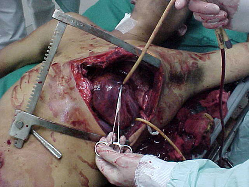
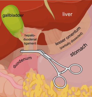
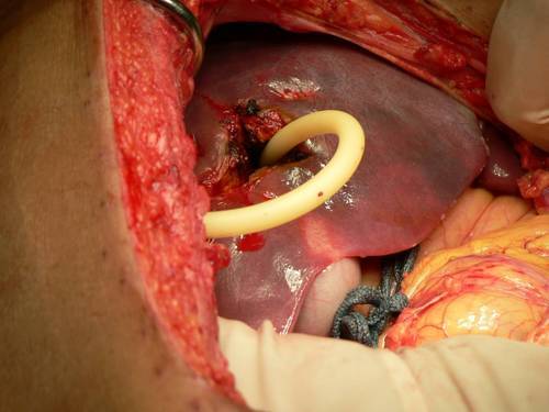
Consider a deep penetrating injury to the liver. It takes time to determine which method for slowing/stopping the bleeding is most appropriate. Sure, the doctor books say to occlude the inflow by gently clamping the hepatoduodenal ligament (Pringle maneuver). But this takes time, and can be difficult if there is lots of bleeding. 

You may be able to gain some time by placing a properly sized Foley catheter directly into the wound and carefully inflating with saline. You must inflate the balloon to feel, not to its full volume. It should be snug, but not so full that it cracks the liver parenchyma and causes yet more bleeding.

Bottom line: Any time you find yourself facing bleeding from hard to expose places, think about using a balloon catheter like the Foley. Sizing is critical, and the balloon volume is more important than the catheter diameter. Estimate the size of the area that needs to be occluded, and then ask for a catheter with a 10cc or 30cc balloon. If you need smaller, more precise control, try a Fogarty arterial embolectomy catheter instead. 

As with the cardiac Foley, be sure to occlude the end so you don’t create a conduit for the blood to escape. If your patient does well, and you need to leave the catheter in place for a damage control closure, LEAVE THE CATHETER COMPLETELY WITHIN THE ABDOMEN. If you exteriorize the end, some well-meaning person may unclamp it, drop the balloon, or decide that it can be used for tube feedings.

TIP: If the distance between the balloon and the catheter tip is too long, DO NOT TRY TO SHORTEN THE TIP BY CUTTING IT! This will damage the balloon and it will not inflate.
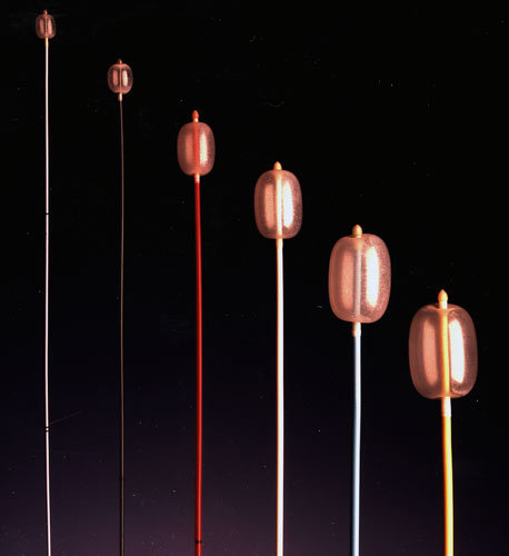
Fogarty catheters

Off-Label Use of the Foley (Urinary) Catheter

Foley catheters are a mainstay of medical care in patients who need control or measurement of urine output. Leave it to trauma surgeons to find warped, new ways to use them!

Use of these catheters to tamponade penetrating cardiac injuries has been recognized for decades (see picture, 2 holes!). Less well appreciated is their use to stop bleeding from other penetrating wounds.

Foley catheters can be inserted into just about any small penetrating wound with bleeding that does not respond to direct pressure. (Remember, direct pressure is applied by one or two fingers only, with no flat dressings underneath to diffuse the pressure). Arterial bleeding, venous bleeding or both can be controlled with this technique. 

In general, the largest catheter with the largest possible balloon should be selected. It is then inserted directly into the wound until the entire balloon is inside the body. Inflate the balloon using saline until firm resistance is encounted, and the bleeding hopefully stops. Important: be sure to clamp the end of the catheter so the bleeding doesn’t find the easy way out!

Use of catheter tamponade buys some time, but these patients need to be in the OR. In general, once other life threatening issues are dealt with in the resuscitation room, the patient should be moved directly to the operating room. In rare cases, an angiogram may be needed to help determine the type of repair. However, in the vast majority of cases, the surgeon will know exactly where the injury is and further study is not needed. The catheter is then prepped along with most of the patient so that the operative repair can be completed.

Related posts:

Trends In Resident Trauma Operative Experience

Even though it’s called trauma surgery, the operative experience in trauma tends to be somewhat limited. This is due mostly to the fact that most trauma centers see predominantly blunt trauma. Yes, there are hospitals around the world where the penetrating injury load remains high and there is operative experience aplenty.

But in the US, the vast majority of trauma centers see mostly blunt trauma. Surgical residents in the US are required to log 10 operative and 20 nonoperative cases to successfully meet residency completion requirements. And blunt trauma is tending to get less and less operative in nature. A good example is the evolution of blunt solid organ injury to mostly nonoperative management.

So what is happening with surgical resident operative trauma experience? And has there been any impact from the work hour restrictions that have gone into effect in the US? A study from Harborview, Denver Health and Seattle Children’s looked at the ACGME operative logs for surgical residents annually from 1989 to 2010. They combined the data into 5 year blocks, with the last two having work hour restrictions in place.

Some interesting findings:

  • Overall mean caseload of major cases (all types) remained steady at about 925 per resident
  • Mean trauma operative caseload decreased from 76 to 39 (beginning of work hour restrictions)
  • Mean trauma operative caseload remained steady at 39 for the 7 years in which work hour restrictions were in effect
  • The number of intra-abdominal trauma operations decreased from 31 to 17, and the number of liver/spleen operations decreased from 5 and 4 to 3 and 2

Bottom line: Resident trauma operative experience has declined and stabilized in the US. This is due to the evolution of our management of blunt trauma. Unfortunately, this decline will reflect on how well prepared surgeons at outlying hospitals are, and in the quality of emergency surgery they may provide. The impact will be felt most by seriously injured patients who cannot be taken to a high level trauma center initially. We need creative solutions to address this issue, such as mini-clerkships in trauma or structured experiences at high level trauma centers for surgeons in outlying hospitals.

Related post: ED at the busiest hospital in the world!

Reference: ACGME case logs: surgery resident experience in operative trauma for two decades. J Trauma 73(6):1500-1506, 2012.

Dysphagia and Cervical Spine Injury

Cervical spine injury presents a host of problems, but one of the least appreciated ones is dysphagia. Many clinicians don’t even think of it, but it is a relatively common problem, especially in the elderly. Swallowing difficulties may arise for several reasons:

  • Prevertebral soft tissue swelling may occur with high cervical spine injuries, leading to changes in the architecture of the posterior pharynx
  • Rigid cervical collars, such as the Miami J and Aspen, and halo vests all force the neck into a neutral position. Elderly patients may have a natural kyphosis, and this change in positioning may interfere with swallowing. Try extending your neck by about 30 degrees and see how much more difficult it is to swallow.
  • Patients with cervical fractures more commonly need a tracheostomy for ventilatory support and/or have a head injury, and these are well known culprits in dysphagia

A study in the Jan 2011 Journal of Trauma outlines the dysphagia problem seen with placement of a halo vest. They studied a series of 79 of their patients who were treated with a halo. A full 66% had problems with their swallowing evaluation. This problem was associated with a significantly longer ICU stay and a somewhat longer overall hospital stay.

Bottom line: Suspect dysphagia in all patients with cervical fractures, especially the elderly. Carry out a formal swallowing evaluation, and adjust the collar or halo if appropriate. 

Reference: Swallowing dysfunction in trauma patients with cervical spine fractures treated with halo-vest fixation. J Trauma 70(1):46-50, 2011.

MRI And External Fixators

MRI is an indispensable tool for evaluation of spine and soft tissue trauma. However, a great deal of effort was be made to ensure that any patient scheduled for this test is “MRI compatible.” The fear is that any retained metallic fragments may move or heat up once the magnets are activated. 

But what about trauma patients with external fixators? That is one big hunk of metal that is inserted deep into your patient. There are three major concerns:

  • Is the material ferromagnetic? If so, it will move when the magnets are activated and may cause internal injury. These days, there are many fixator sets that are not ferromagnetic, avoiding this problem.
  • Can currents be induced in the material, causing heating? This is not much of a problem for small, isolated objects. However, external fixators are configured in such a way that loops are created. The fluctuating magnetic fields can induce currents that in turn will heat the surrounding tissue. And thinner materials (narrow pins) result in more current and more heating.
  • Will the metal degrade image quality?

The biggest challenge is that there is no standard ex-fix configuration. Our orthopaedic colleagues get to unleash their creativity trying to devise the appropriate architecture to hold bones together so they can heal properly. This makes it difficult to develop standardized guidelines regarding what can and can’t go into the scanner.

However, there is a growing body of literature showing that the heating effects are relatively small, and get smaller as the distance from the magnet increases. And non-ferromagnetic materials move very little, if at all, and do not interfere with the image. So as long as nonferromagnetic materials are used, the patients are probably safe as long as basic principles are adhered to:

  • Other diagnostic options should be exhausted prior to using MRI.
  • Informed consent must be obtained, explaining that the potential risks are not completely understood.
  • The fixator must be tested with a handheld magnet so that all ferromagnetic components can be identified and removed.
  • All traction bows must be removed.
  • Ice bags are placed at all skin-pin interfaces.
  • The external fixator must remain at least 7cm outside the bore at all times.

Bottom line: MRI of patients with external fixators can be safely accomplished. Consult your radiologists and physicists to develop a policy that is specific to the scanners used at your hospital. 

Related posts: