All posts by The Trauma Pro

Penetrating Injuries to the Extremities

Simple penetrating injuries to the arms and legs are often over-treated with invasive testing and admission for observation. Frequently, these injuries can be rapidly evaluated and disposed of using physical examination skills alone.

Stabs and low velocity gunshots (no rifles or shotguns, please) should be thoroughly examined. This includes an examination of the entire, unclothed body. If this is not carried out, there is a risk that additional penetrating injuries may be missed.

For gunshots, look at the wounds and the estimated trajectory to try to demonstrate that the object stayed clear of neurovascular structures. This exam is imprecise, and must be accompanied by a full neurovascular exam and evaluation of the bones and joints. If there is any doubt regarding bony involvement, plain radiographs with entry markers should be performed. Any abnormal findings will require more in-depth evaluation and inpatient admission.

If the exam is negative but the trajectory is “in proximity” to a major vessel, an arterial pressure index (API) should be measured. This test involves the calculation of the ratio of the systolic pressure in the injured extremity to the contralateral uninjured extremity. It should not be confused with the ankle brachial index (ABI) which compares the systolic pressure in the ipsilateral uninjured arm  or leg.

The magic ratio is 0.9. If the API is less than this, there is some likelihood that a vascular injury is present. If the API is higher, there is virtually no chance of injury.

The final test that must be performed before discharge is a function test. If the injured extremity is too painful to use or walk on, the patient may need to be admitted for pain management and therapy. Patients managed in this way can avoid arteriography, CT angiography or admission and save thousands of dollars in hospital charges.

Reference: Journal Am Coll Surgeons 2009;209:740-5.

A Better Concussion Test?

Concussions from sports activities are all over the news these days. This injury is particularly important when dealing with the developing brains of student athletes. Most organized sporting activities take advantage of existing concussion testing on the sidelines, such as the Standardized Assessment of Concussion test (SAC, a cognitive test), and the tandem gait test (balance). 

However, sports concussions frequently involve the visual pathways as well. In an effort to improve concussion recognition, a New York group studied the addition of a visual test (King-Devick test, K-D) to their concussion battery. A large group of youth and collegiate hockey and lacrosse players were evaluated at their baseline with K-D, SAC, and tandem gait. During games, athletes with potential concussions were tested on the sidelines. They were compared to non-concussed athletes who were also tested on the sidelines.

Here are the factoids:

  • 243 student athletes were studied, average age 11
  • 12 athletes sustained concussions, and their performance on the K-D test worsened significantly
  • The K-D test identified concussions better than the cognitive and balance tests

Bottom line: The addition of the King-Devick test appears to improve concussion identification in young athletes. In fact, it may be possible to use just this test alone. But practically speaking, it is probably better to use as part of a battery of tests to identify possible concussion after a significant impact. This enables coaches to minimize the number of players that might sustain additional injury and develop the risk for later sequelae of concussion. 

Related posts:

Reference: Adding vision to concussion testing: a prospective study of sideline testing in youth and collegiate athletes. J Neuro-opthalmology 2015 Feb 18, epub ahead of print.

Pigtail Catheters Instead Of Chest Tubes?

Traditionally, hemothorax and pneumothorax in trauma has been treated with chest tubes. I’ve previously written about some of the debate regarding using smaller tubes or catheters. A paper that will be presented at the EAST meeting in January looked at pain and failure rates using 14Fr pigtail catheters vs 28Fr chest tubes.

This was a relatively small, prospective study, and only 40 of 74 eligible patients were actually enrolled over 20 months at a Level I trauma center in the US. Pain was measured using a standard Visual Analog Scale, as was complication and failure rate, tube duration and hospital stay.

The following interesting findings were noted:

  • Chest wall pain was similar. This is expected because the underlying cause of the pneumothorax, most likely rib fractures, is unchanged.
  • Tube site pain was significantly less with the pigtail
  • The failure rate was the same (5-10%)
  • Complication rate was also the same (10%)
  • Time that the tube was in, and hospital stay was the same

Bottom line: There may be some benefit in terms of tube site pain when using a smaller catheter instead of a chest tube. But remember, this is a very small study, so be prepared for different results if you try it for your own trauma program. If you do choose to use a smaller tube or catheter, remember to do so only in patients with a pure pneumothorax. Clotted blood from a hemothorax will not be completely evacuated.

Related posts:

Reference: A prospective randomized study of 14-French pigtail catheters vs 28F chest tubes in patients with traumatic pneumothorax: impact on tube-site pain and failure rate. EAST Annual Surgical Assembly, Oral paper 12, 2013.

Trauma MedEd Newsletter Released To Subscribers Tonight!

The March Trauma MedEd Newsletter will be released to subscribers tonight. This month’s theme is “the elderly.” Articles include:

  • How we take care of our elders
  • Falls, falls, and more falls
  • The need for a medical orthopedic service
  • And some philosophy on geriatric trauma

Anyone on the subscriber list as of noon (CST) today will receive it tonight. I’ll release it to everyone else next Monday via the blog. So sign up for early delivery now by clicking here!

Pick up back issues here!

Do You Really Need To Repeat That Xray?

It happens all the time. You get that initial chest and/or pelvic xray in the resuscitation room while evaluating a blunt trauma patient. A few minutes later the tech returns with another armful of xray plates to repeat them. Why? The patient was not centered properly and part of the image is clipped.

Do you really need to go through the process of setting up again, moving the xray unit in, watching people run out of the room (if they are not wearing lead, and see my post below about how much radiation they are really exposed to), and shooting another image? The answer to the question lies in what you are looking for. Let’s address the two most common (and really the only necessary) images needed during early resuscitation of blunt trauma.

First, the chest xray. You are really looking for 3 things:

  • Big air (pneumothorax)
  • Big blood (hemothorax)
  • Big mediastinum (hinting at aortic injury)

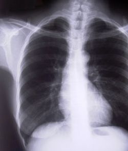
Look at the clipped xray above. A portion of the left chest wall is off the image. If there were a large pneumothorax on the left, would you be able to see it? What about a large hemothorax? And the mediastinum is fully included, so no problem there. So in this case, no need to repeat immediately.

The same thing goes for the pelvis. You are looking for gross disruption of the pelvic ring, especially posteriorly because this will cause you to intervene in the ED (order blood, consider wrapping the pelvis). So if parts of the edges or top and bottom are clipped, no big deal.

Bottom line: Don’t let the xray tech disrupt the team again by reflexively repeating images that are not technically perfect. See if you can use what you already have.  And how do you decide if you need to repeat it later, if at all? Consider the mechanism of injury and the physical exam. Then ask yourself if there is anything you could possibly see that was not imaged the first time that would change your management in any way. If not, you don’t need it. But it certainly will irritate the radiologists!

Related posts: