All posts by The Trauma Pro

March Trauma MedEd Newsletter Is Coming!

Finally! After a bit of a delay, the issue on REBOA is being released to subscribers this weekend. Here is what you will find inside:

  • Everything You Always Wanted To Know About REBOA
  • What Is REBOA?
  • Who is REBOA For?
  • How Is REBOA Performed?
  • What Are The Results For REBOA?
  • What’s The Bottom Line?

Subscribers will receive it over the weekend; everyone else will have to wait until the end of next week.

Subscribe now and be sure to get it first!  So sign up for early delivery now by clicking here!

Pick up back issues here!

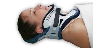
Comparison of Cervical Spine Stabilization

A reader recently asked what the optimal method for inline stabilization is. We’ve been pondering this question for nearly 30 years. In 1983, trauma surgeons at UCLA looked at a number of devices available at that time and tested them on normal volunteers. They measured neck motion to see which was “best.”

Here’s what they found:

  • Soft collar – In general, this decreased rotation by 8 degrees but insignificantly protected against flexion and extension. Basically, this keeps your neck warm and little else.
  • Hard collars – A variety of collars available in that era were tested. They all allowed about 8% flexion, 18% lateral movement, and 2% rotation. The Philadelphia collar allowed the least extension.
  • Sandbags and tape – Surprisingly, this was the best. It allowed no flexion and only a few percent movement in any other direction.

The Mayo clinic compared four specific hard collars in 2007 (Miami J, Miami J with Occian back, Aspen, Philadelphia). They found that the Miami J and Philadelphia collars reduced neck movement the best. The Miami J with or without the Occian back provided the best relief from pressure. The Aspen allowed more movement in all axes.

And finally, the halo vest is the gold standard. These tend to be used rarely and in very special circumstances.

Bottom line:

  • For EMS: Rigid collar per your protocol is the standard. In a pinch you can use good old tape and sandbags with excellent results.
  • For physicians: The Miami J provides the most limitation of movement. If the collar will be needed for more than a short time, consider the well-padded Occian back Miami J (see below).

References:

  • Efficacy of cervical spine immobilization methods. J Trauma 23(6):461-465, 1983.
  • Range-of-motion restriction and craniofacial tissue-interface pressure from four cervical collars. J Trauma 63(5):1120, 1126, 2007.

Falls vs Abuse In Kids: Differences In Injury Patterns

Nonaccidental trauma (NAT) in children, a.k.a. child abuse, is a problem that trauma professionals see all too frequently. Much of the time, the abuse is obvious. Sometimes, it is more insidious and occult, and we can be misdirected by the history given by the caregivers. The most frequent story used to cover up obvious injuries child abuse is that the child fell. Unfortunately, the injuries observed from abuse may be very similar to those seen from shaking, grabbing, lifting, and throwing.

A paper that is currently in press from the University of Colorado at Aurora seeks to clarify this a bit, trying to tease out nuances in common injury patterns that may help us distinguish NAT from falls. They performed a retrospective database review at both Denver Health and Children’s Health Colorado over a 15 year period. They specifically looked at children with blunt abdominal trauma. Unfortunately, they chose the age group < 18 years as “children”, which muddies the picture somewhat. 

Here are the factoids:

  • Of the 1,005 blunt abdominal trauma cases identified, 65 were confirmed to be due to NAT, and 115 were actually from falls
  • 63 of the 65 NAT victims were less than 5 years old, but only 35 of the falls were
  • Average ISS for the NAT kids was 20, vs only 12 for falls
  • There were more hollow viscus injuries in NAT kids (25 vs 2), and more pancreatic injuries (16 vs 2)
  • If a head injury was present, it was more severe with NAT
  • Hospital LOS was longer after NAT, which makes sense given the ISS and head info above

Bottom line: Unfortunately, the authors could accumulate only a small amount of data over 15 years, but it paints a clear picture. Injured children presenting with a history of falls, particularly young children who can’t engage in the high energy pursuits of adolescents, should arouse suspicion. If multiple injuries are found, especially visceral or deep solid organ abdominal injury (pancreas), suspect foul play. Similarly, if the head injury is more severe, be suspicious. All trauma professionals need to keep the possibility of NAT in the back of their minds on every injured child they see!

Related posts:

Reference: Pediatric abdominal injury patterns caused by “falls”: A comparison between nonaccidental and accidental trauma. J Ped Surg, in press, Feb 2, 2016.
Impact Of Arm Position On Torso CT Scan

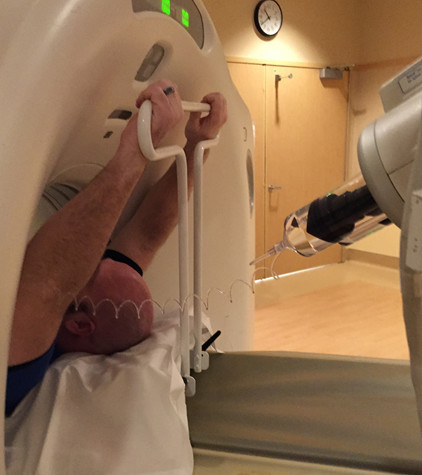
CT scan is a valuable tool for initial screening and diagnosis of trauma patients. However, more attention is being paid to radiation exposure and dosing. Besides selecting patients carefully and striving for ALARA radiation dosing (as low as reasonably achievable) by adjusting technique, what else can be done? Obviously, shielding parts of the body that do not need imaging is simple and effective. But what about simply changing body position?

One simple item to consider is arm positioning in torso scanning. There are no consistent recommendations for use in trauma scanning. Patients with arm and shoulder injuries generally keep the affected upper extremity at their side. Radiologists prefer to have the arms up if possible to reduce scatter and provide clearer imaging.

A retrospective review of 710 patients used dose information computed by the CT software and displayed on the console. Radiation exposure was estimated using this data and was stratified by arm positioning. Even though there are some issues with study design, the results were impressive.

There was no difference in scanning time for any arm position. Here are the factoids for radiation dose:

  • Both arms up: 19 mSv (p<0.0000001)
  • Left arm up: 23 mSv
  • Right arm up: 24 mSv
  • Arms down: 25 mSv

Bottom line: Do everything you can to reduce radiation exposure:

  1. Be selective with your imaging. Do you really need it?
  2. Work with your radiologists and physicists to use techniques that reduce dose yet retain image quality
  3. Shield everything that’s not being imaged.
  4. Think hard about getting CT scans in children. They probably don’t need it!
  5. Raise both arms up during torso scanning unless injuries preclude it.

There is a commercial product now available that helps position the arms without tape, paper clips, or other office supply items. It doubles as a pillow for the patient and is held in place by their weight

Courtesy of http://accessoryaccommodations.com/

Related posts:

Reference: Influence of arm positioning on radiation dose for whole body computed tomography in trauma patients. J Trauma 70(4):900-905, 2011.

Can Lead Poisoning Occur After A Gunshot?

This is a fairly common question from victims of gunshots and their families. As you know, bullets are routinely left in place unless they are superficial. It may cause more damage to try to extract one, especially if it has come to rest in a deep location. But is there danger in leaving the bullet alone?

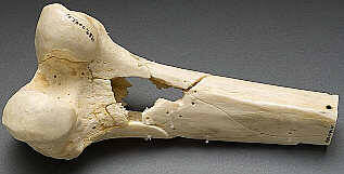
One of the classic papers on this topic was published in 1982 by Erwin Thal at Parkland Hospital in Dallas. The paper recounted a series of 16 patients who had developed signs and symptoms of lead poisoning (plumbism) after a gunshot or shotgun injury. The common thread in these cases was that the injury involved a joint or bursa near a joint. In some cases the missile passed through the joint/bursa but came to rest nearby, and a synovial pseudocyst formed which included the piece of lead. The joint fluid bathing the projectile caused lead to leach into the circulation.

The patients in the Parkland paper developed symptoms anywhere from 3 days to 40 years after injury. As is the case with plumbism, symptoms were variable and nonspecific. Patients presented with abdominal pain, anemia, cognitive problems, renal dysfunction and seizures to name a few.

Bottom line: Any patient with a bullet or lead shot that is located in or near a joint or bursa should have the missile(s) promptly and surgically removed. Any lead that has come to rest within the GI tract (particularly the stomach) must be removed as well. If a patient presents with odd symptoms and has a history of a retained bullet, obtain a toxicology consult and begin a workup for lead poisoning. If levels are elevated, the missile must be extracted. Chelation therapy should be started preop because manipulation of the site may further increase lead levels. The missile and any stained tissues or pseudocyst must be removed in their entirety.

Reference: Lead poisoning from retained bullets. Ann Surg 195(3):305-313, 1982.