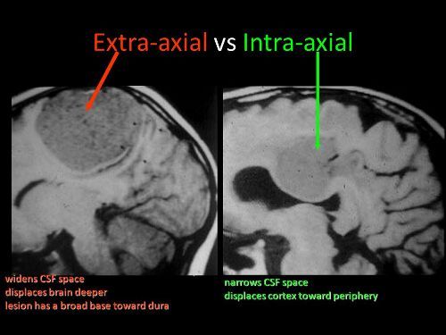
You’ve seen it on head CT reports. “The patient has a collection of extra-axial blood…” Then it goes on to describe the location and size of a subdural hematoma. But why is it called “extra-axial?”
The answer lies in the embryology of the central nervous system. Yes, it’s been a long time since any of us have read anything about that. Early animals had a straight neural tube, which slowly evolved into a brain and spinal cord. This is known as the axis of the nervous system.
The brains of early vertebrates developed at the end of the neural tube, and were oriented in the same longitudinal axis as the rest of it. As brains got bigger, a 90 degree bend occurred at the cephalic flexure.
So in humans, there is a difference between the body axis and the brain axis. But the brain axis is what really counts. This means that any blood outside of the brain axis is defined as extra-axial.
Bottom line: Extra-axial blood is defined as any bleeding outside of the brain parenchyma. This includes subdural and epidural hematomas, and subarachnoid hemorrhage. It does not include any intraparenchymal bleeding like contusions, strokes, or hematomas.
Yesterday, I wrote about proper screening for blunt cerebrovascular injury (BCVI). But, as you know, it’s important to screen only when there is a significant risk of the injury being present. Screening using the shotgun approach (screen everyone for everything) yields enough false positive results to present potential danger to your patient.
A variety of authors on this topic have promoted a number of high risk criteria to trigger a screening test. Most make sense, and are related to the anatomy of the vessels in question. The carotid arteries are relatively unprotected, although a bit deep, as they course up the neck. Thus, it is possible to damage them when they suffer a direct and significantly hard blow. Once they enter the skull, they are better protected. However, fractures through key areas of the skull base and face can injure the vessels, even in these protected locations.
The vertebral arteries are deep and relatively protected as they course through the vertebral foramina. However, if the vertebrae are fractured or subluxed, vessel injury can occur.
Finally, and as always, the physical exam is important. If there are unexpected neurologic changes that can’t be explained by other injuries, or there are indications of deep vascular injury, BCVI needs to be considered.
Here is my list of indications to screen for BCVI:
Neurologic abnormality not explained by diagnosed injury
Arterial epistaxis
Seat belt sign on neck
GCS < 8 (this is the most commonly forgotten one)
Petrous bone fracture
C‐spine fracture (C1‐C3) or subluxation at any level
Fracture through foramen transversum
LeFort II or III fractures
Bottom line: Be on the lookout for any of the criteria listed above in your trauma patient. If you find one during your initial evaluation, be sure to order a CT angiogram of the neck. And keep an eye out while scanning the head and cervical spine. If any of the other radiographic indications become apparent, add on the CT angiogram at that point.
Blunt injury to the carotids or vertebrals (BCVI) is a little more common than originally thought, affecting about 1% of blunt trauma patients. We have many tools available to help us diagnose the problem: duplex ultrasound, CT angiography (CTA), MR angiography (MRA), and even good old conventional 4 vessel angiography.
But which one is “best?” This is a tough question, because there is always some interplay between clinical accuracy and cost. The surgical group at the Medical College of Wisconsin – Milwaukee did a nice job teasing some answers from existing literature on the topic. The authors tried to take a comprehensive look at costs, including money spent to prevent stroke, the cost of complications of therapy, and the overall cost to society if the patient suffers a stroke.
Here are the factoids:
For patients at risk for BCVI, the stroke rate is 11% without screening, 6% with duplex ultrasound screening, 4% with MRA, and 1% with either CTA or conventional angiography
From a societal standpoint (includes the lifetime costs of stroke for the patient), CTA is the most cost effective at $3,727 per patient
From the hospital standpoint (does not include lifetime cost), no screening is the most cost effective, but has the highest stroke rate (11%)
CTA prevents the most strokes, and costs about $10,000 per patient while decreasing societal costs by about $32,000 per patient screened
Bottom line: The “best” test for patients at risk for blunt cerebrovascular injury is the CT angiogram. It minimzes the stroke rate, and provides information on all four vessels supplying the brain, which is probably why the duplex ultrasound has a higher miss rate (can’t see the vertebrals or into the skull). But how do you decide who is at risk for this problem. Tune in tomorrow!
Reference: Screening for Blunt Cerebrovascular Injuries is Cost-Effective. J Trauma 70(5):1051-1057, 2011.
With the implementation of resident work hour restrictions 10 years ago, resident participation in clinical care has declined. In order to make up for this loss of clinical manpower and expertise, many hospitals have added advanced clinical providers (ACPs, nurse practitioners and physician assistants). These ACPs are being given more and more advanced responsibilities, in all clinical settings. This includes performing invasive procedures on critically ill patients.
A recent study from Carolinas Medical Center in Charlotte NC compared complication rates for invasive procedures performed by ACPs vs residents in a Level I trauma center setting.
A one year retrospective study was carried out. Here are the factoids:
Residents were either surgery or emergency medicine PGY2s
ACPs and residents underwent an orientation and animal- or simulation-based training in procedures
All procedures were supervised by an attending physician
Arterial lines, central venous lines, chest tubes, percutaneous endoscopic gastrostomy, tracheostomy, and broncho-alveolar lavage performances were studied
Residents performed 1020 procedures and had 21 complications (2%)
ACPs performed 555 procedures and had 11 complications (2%)
ICU and hospital length of stay, and mortality rates were no different between the groups
Bottom line: Resident and ACP performance of invasive procedures is comparable. As residents become less available for these procedures, ACPs can (and will) be hired to take their place. Although this is great news for hospitals that need manpower to assist their surgeons and emergency physicians, it should be another wakeup call for training programs and educators to show that resident education will continue to degrade.
Reference: Comparison of procedural complications between resident physicians and advanced clinical providers. J Trauma 77(1):143-147, 2014.
Yesterday, my colleague the Skeptical Scalpel wrote about an interesting (?) paper published in Emergency Medicine Australasia. It was a small study that concluded that ED wait times decreased as the number of people presenting to be seen decreased. Where’s the mystery in that? Overstating the obvious?
But if you look through almost any journal today, you will find studies that leave you wondering how they ever got published. And this is not a new phenomenon. Look at any journal a year ago. Five years ago. Twenty years ago. And even older. The research landscape is littered with their carcasses.
And on a related note, sit down with any serious clinical question in your field you want to answer. Do a deep dive with one of the major search engines and try to get an answer. Or better yet, let the professionals from the Cochrane Library or other organization do it for you. Invariably, you will find hints and pieces of the answer you seek. But never the completely usable solution you desire.
Why is it so hard? With tens of thousands of articles being published every year?
Because there is no plan! Individuals are forced to produce research as a condition of their employment. Or to assure career advancement. Or to get into medical school, or a “good” residency. And in the US, Level I trauma centers are required to publish at least 20 papers every three years to maintain their status. So there is tremendous pressure across all disciplines to publish something.
Unfortunately, that something is usually work that is easily conceived and quickly executed. A registry review, or some other type of retrospective study. They are easy to get approval for, take little time to complete and analyze, and have the potential to get published quickly.
But what this “publish or perish” mentality promotes is a random jumble of answers that we didn’t really need. There is no planning. There is no consideration of what questions we really need to answer. Just a random bunch of easy to get published thoughts that never get cited by anyone else.
Bottom line: How do we fix this? Not easily. Instead of focusing on the quantity of publications, the “authorities” need to focus in on their quality. Extra credit should be given to multicenter trial involvement, prospective studies, and other higher quality projects. The actual number of publications should not matter as much as how much high quality work is in progress. Sure, the sheer number of studies published will decline, but the quality will increase exponentially!
Home of the Trauma Professional's Blog
Do you want to get a daily email every time there’s a new post? See what I’m up to.