All posts by The Trauma Pro

REBOA Part 2: Who Will Benefit?

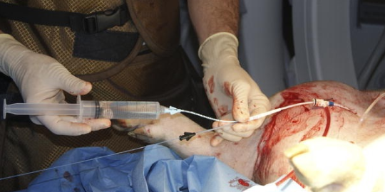
First, I would like to state that REBOA is not for the faint of heart. Hmm, not a very good idiom. It actually might be, if you are the patient.

I say this because REBOA has a definite learning curve from a technical standpoint. But it does use standard trauma and vascular surgical techniques, which makes it a little easier to grasp. At this point, it should primarily be performed by surgeons, since it frequently creates a vascular injury that requires surgical repair at the end of the procedure. However, to be fair, emergency physicians can and do initiate the procedure here and in some countries outside the US, such as Japan. Terminating it is another matter.

From a patient selection standpoint, think of it as a way of keeping your patient alive until you can get them to the OR for definitive control of their hemorrhage. You are trading 5 to 10 more minutes in the trauma bay inserting it for a (potentially) safer trip to the OR suite, and lets the surgeons start the case with some modicum of vascular control already in place.

The abdomen is divided into 3 REBOA zones, depending on where the hemorrhage is located. Here’s the map:

image

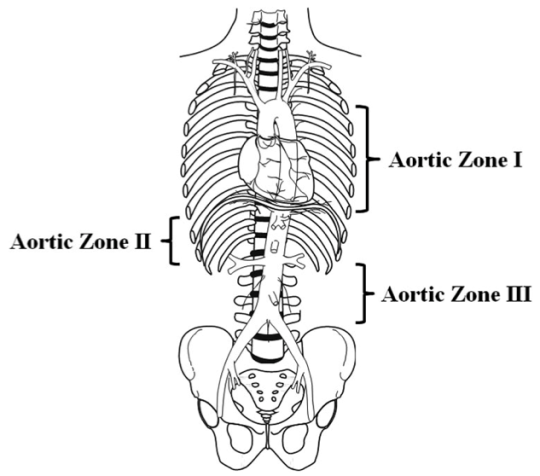
For bleeding in the abdominal cavity, the REBOA balloon is placed in Zone I. For practical purposes, we try to occlude the distal aorta at the diaphragm, where we would normally place the crossclamp for an ED thoracotomy.

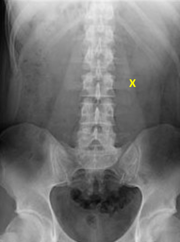
For pelvic bleeding, generally from branches of the iliac arteries, the balloon is placed in the distal aorta, Zone III. Zone II is not used currently.

So who will benefit from REBOA? The answers to this question are still being teased out of the small series that are being produced by a number of centers. The general rule is that any patient with exsanguinating hemorrhage originating below the diaphragm should be considered for this procedure.

Does that mean all patients? Patients who still have vital signs? How good or bad do they need to be? Unfortunately, we don’t know yet. But we are working on it.

Monday: How is REBOA performed?

Direct links to the REBOA series:

REBOA Part 1: What Is It?

Technically, REBOA is the acronym for Resuscitative Endovascular Balloon Occlusion of the Aorta. It is a relatively new tool in our armamentarium for use in patients with uncontrolled hemorrhage. Essentially, it allows the surgeon to crossclamp the aorta at just about any level, without opening the chest or abdomen.

But as with anything new, it is usually derived from something old. And REBOA is no exception. Case reports surfaced in the Korean war, and continued through the 1980s. The technique was then adopted by vascular surgeons and used for controlling hemorrhage above a ruptured abdominal aortic aneurysm. As with most major trauma “discoveries”, military conflict also tends to foster the development of new and the refinement of existing techniques.

The early part of this decade was actually the heyday for animal testing of this technique. Numerous pigs were sacrificed in order to show that 1) it could be done relatively safely, 2) it definitely increased blood flow to the brain and heart, and 3) it decreased mortality. Finally, the technique was shown to have similar effects and outcomes to pig thoracotomy with cross-clamping.

image

The first small human series was published just a year ago, so our experience is relatively short and limited to small series. But it continues to grow steadily, and more and more trauma centers are beginning to dabble with the technique.

Tomorrow: Who would benefit from REBOA?

Direct links to the REBOA series:

Everything You Always Wanted To Know About: REBOA!

REBOA has become one of the hot topics that everyone seems to be talking about (and writing about). As with any hot new trend, it’s important to understand the facts, as much as they’ve been worked out. The enthusiasts are, by definition, always very enthusiastic, and sometimes the hype overshadows the reality.

During the next week, I’m going to methodically make my way through the basics, like what it is, how we came up with the idea, and what it entails. Then I’ll look through the literature as we know it. Finally, I’ll try to put it all together and make some recommendations about what you should be doing with it.

Tune in, starting Tomorrow!

Direct links to the REBOA series:

Now Where Did That Bullet Go? The Answer!

Previously, I presented a scenario where a victim of a gunshot to the abdomen was taken to the OR after obtaining the image below. No bullet was seen on the x-ray, and none was found at the time operation.

image

Where could it have gone? Let’s assume that the surgeon did a good job, and it is not in the abdomen. Any more. There are several possibilities.

Does the x-ray cover the correct area? To cover a straighforward abdominal gunshot, it needs to show diaphragms to perineum, side to side. In these days of “super size me”, that usually doesn’t happen with one image. Look carefully at the one above. It doesn’t show any portion of the diaphragm, and doesn’t go low enough, either. And although the right flank can be seen, the left is cut off. So in this case, the bullet could be in the soft tissues of the torso, in the extraperitoneal rectal area, or near the diaphragm in the liver.

It could have moved outside the area of the initial x-ray. The most common mechanism for this is entry into the vascular system. If it enters the venous system, it will end up in the heart or pulmonary artery somewhere. This will be obvious when you get a chest x-ray. If it enters the aorta, it will embolize into the lower extremities. This fact should be painfully obvious when you check the pulses in the lower extremities

The patient could poop it out if it entered their GI tract. This could happen if you wait to get additional images of the abdomen. If you bracket it with x-rays immediately, this should not happen. 

In theory, the bullet could enter the bladder and get urinated out. This won’t happen if a catheter is in place. And it’s probably unrealistic because most bullets would cause tremendous pain passing, and would probably obstruct the urethra anyway.

Finally, it could have bounced. Never count on this one. Bullets can and do enter partially, then stop or fall out. They can cause underlying perforation of the peritoneum, and they can bruise nearby structures. This is extremely uncommon and should be a diagnosis of last resort!

Bottom line: If patient condition permits, the patient with a gunshot to the abdomen who will be taken to the OR should have any wounds marked and an initial abdominal image obtained that shows the entire abdomen. This may take multiple attempts. The image can be very helpful in directing the exploration and finding wounds. If it is not seen on the initial image(s), check the lower extremity pulses and obtain a chest x-ray to locate the bullet prior to the case.

Now Where Did That Bullet Go?

Here’s an interesting case for you to consider. A male victim of an assault is brought to your emergency department with a gunshot to the abdomen. He is met by your team as a trauma activation. Vitals are stable, but he has guarding and rigidity. A single abdominal x-ray is obtained, and then the patient is taken off to surgery. I’ve marked the entry site on the anterior abdominal wall below. There is no exit wound.

In the operating room a laparotomy is performed. There is a hole in the fascia under the skin penetration. A small hematoma is seen in the underlying transverse mesocolon, well away from the bowel itself. An extensive search is carried out, but no other holes, injuries, or evidence of a bullet is found. Ultimately, the abdomen is closed and the patient is admitted to a ward bed.

WTF? Where did the bullet go? What do you think the possibilities are? Please leave comments today either here or on Twitter. I’ll analyze this puzzling situation tomorrow!

Disclosure for my social media compliance police: this patient was not treated at Regions Hospital, and the x-ray was obtained and modified from the internet.