All posts by The Trauma Pro

Trauma In Pregnancy 4: Imaging

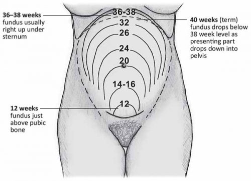
Everyone worries about imaging pregnant patients. As with most medical tests, it always boils down to risks vs benefits. What are the chances of causing mutations or cancers or a spontaneous abortion, and what is the risk of missing a critical injury? In general, reasonable studies involving a fetus at just about any point in gestation won’t cause major problems. At least as far as we know. What is not clear are the longer term, hard to measure effects. So the general philosophy should be to order just what you absolutely need, and shield the fetus during any studies other than of the abdomen/pelvis.

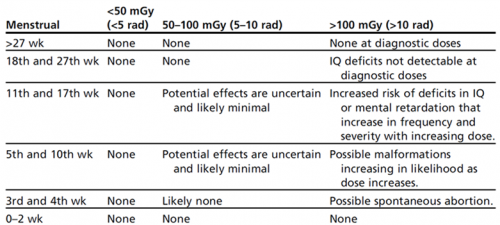
Now, to put these numbers into perspective, have a look at this list of delivered doses from common studies. The table above is listed in milliGrays, and this one is in milliSieverts. These are roughly comparable, except that the former is a measure of radiation dose absorbed, and the latter measures radiation delivered.

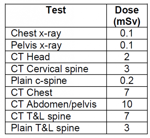
Bottom line: Think hard about the imaging you really need. If you generally do this for all patients, you probably won’t change your practice in pregnant women. Don’t worry about chest and pelvic x-rays. Shield the fetus for anything not involving the abdomen/pelvis. For major torso trauma, you probably will need CT of the chest/abdomen/pelvis. If so, do it right. Order with contrast so you don’t get substandard images that need to be repeated.

Trauma In Pregnancy 3: Tips & Tricks

Here is a list of practical tips for trauma professionals from prehospital and beyond:

  • Apply supplemental oxygen from the get-go, in the field if possible
  • Start an IV or IO and begin crystalloid infusion immediately
  • Bump the patient to her left. This is most easily done with the spine board in place. Just put a rolled up towel under the right side of the board.
  • Quickly estimate how pregnant the patient is if she can’t tell you. Use the fundal height diagram below. Key measurements to remember:
    • Umbilicus = 20 weeks: entering the possible viability range if delivered emergently
    • Xiphoid = approaching full term

  • If the patient is 20 weeks+ pregnant, only transport to a trauma center (Level I or II).
  • At the hospital, reassess fundal height to more accurately estimate viability
  • If the fetus may be viable (23-24 weeks), call OB to the resuscitation area. But remember, they have nothing to offer until the trauma team secures the safety and stability of the mother. So don’t let them in the room until you have finished your entire evaluation.  Nothing they can do (e.g. monitor) is helpful during the acute phase of the resuscitation.
  • Once the mother is stabilized, assess fetal heart tones and apply monitors for fetal heart rate and maternal contractions.
  • Add coag studies to your lab panel. Fetal and placental injury can cause clotting problems.
  • Consider giving Rhogam in all major trauma cases if the mother is Rh negative
  • Obtain a KB test to look at the amount of fetal blood in the maternal circulation. Why do this if you are going to give Rhogam anyway? Answer: sometimes the amount of fetal blood leakage is greater than that covered by a single vial of Rhogam.
  • Plan your imaging intelligently. The next section covers this topic in more detail.
  • If needed, chest tubes should be inserted 1-2 intercostal spaces higher than usual.
  • The life-saving tetanus shot is safe in pregnant patients.


Trauma In Pregnancy 2: Predicting Outcome

The data on maternal outcome after trauma is mixed and somewhat confusing. Mortality after major trauma actually appears to be less. However, injury severity score (ISS) still correlates fairly well with overall mortality. But interestingly, other outcomes (complications) appear to be worse, even for relatively minor injuries. The reason behind this is not clear. Could it be a result of all of the physiologic changes noted above, hormonal factors, or something we don’t fully understand?

Fetal outcome is a function of the mechanism of injury (blunt vs penetrating), and extreme injury severity in the mother. Penetrating injury is uniformly devastating to the fetus, with 70% mortality for gunshots and 40% for stabs. Fetal death from blunt injury is primarily a function of placental abruption. About two thirds of blunt fetal deaths are due to abruption, with 50% of them due to car crashes. Maternal ISS does not correlate with fetal death, except in cases of very high scores. These women most likely experience anatomic and physiologic injuries that lead to fetal demise.

Tomorrow: Tips & Tricks

Reference: Trauma during pregnancy. OB Clinics of North America 40:47-57, 2013.

Trauma In Pregnancy 1: Introduction

Trauma in the pregnant patient is scary, but thankfully not very common. About 1 in 15 pregnant women sustain some type of significant injury. About half are from car crashes (most commonly if unrestrained), and 25% each are from assault (frequently domestic) and falls. Unfortunately, trauma is the leading cause of non-obstetric fetal death.

What makes this type of trauma problematic is two-fold, literally. First, you have two patients. And second, neither one has “normal” physiology. The mother has made numerous adaptations to accommodate the pregnancy, and the fetus is essentially a small parasite, totally dependent on her.

Let’s look at the impact of some of the more important maternal adaptations:

  • Total blood volume increases by about one liter. This would seem to be good, but since it is mainly dilutional with no real increase in total RBCs, and this is coupled with a lower mean blood pressure. Trauma professionals can easily underestimate blood loss.
  • Going into the third trimester, the uterus can compress the IVC when the mother is lying supine. but it is quite frequently forgotten. One of the first maneuvers when you suspect an advanced pregnancy is to bump the patient to her left. Do this even if the blood pressure is normal.
  • The pelvic veins are huge. Disruption from pelvic fractures or penetrating injury can lead to massive bleeding.
  • The stomach is probably full, and under pressure from below. This increases aspiration risk in women who have decreased mental status or need intubation.

And don’t forget the fetus:

  • The fetal/placental/uterine complex is one large, non-compressible unit with multiple shear planes within it. Blunt force will stress those planes, and may result in disruption of the uterus from pelvic veins (massive bleeding), or separation of the placenta from the uterus (abruption).
  • The fetus is totally dependent on the mother for survival, but the placenta will protect the mother first, shutting down fetal circulation if she becomes hypotensive or hypoxic.
  • The baby was designed to come into this world at full term. We have developed the technology to sustain life in smaller and more premature babies. The magic number of weeks keeps slowly decreasing, but preemie survival without complications is a challenge.

Tomorrow, we’ll move on and get to the fun stuff, predicting outcome after trauma in pregnancy.

Dealing With Trauma Flow Sheet Documentation Problems

Over the years, I’ve commented a number of times on paper vs electronic trauma flow sheet. For those of you who somehow missed it, let me recap. Don’t use an electronic trauma flow sheet yet if you can possibly avoid it!

I look at the flow sheet as having two phases, input and output. The input phase occurs as data is being recorded on the sheet, hopefully in real time as events occur during the trauma resuscitation and its aftermath. The output phase consists of a human reviewing the completed flow sheet and analyzing the events and timing for performance improvement (PI) purposes.

The electronic trauma flow sheet has major problems in both phases. But the good, old-fashioned paper sheet isn’t perfect either. It is subject to problems during the input phase. The most common issue is incomplete documentation. I’ve seen so many trauma programs with ongoing issues in this area, and they struggle to find ways to improve or eliminate the missing data.

Here are a few tips you should consider:

  • Make sure your paper flow sheet is well-designed. Data items should not be scattered randomly over several pages. Primary survey items should be grouped together. Medications must have their own block. Diagnostic tests performed (not ordered) should be in the same area. Make sure that the narrative block that typically has vital signs and free-form text about what is happening is large enough, with enough room to write comfortably. There are so many good trauma flow sheets out there already. Borrow a few to see if your program can adopt some of the organizational concepts found on them.
  • Identify the commonly incomplete items at your program, then redesign the flow sheet to cluster them together in one prominent spot on it. Common missed items include patient temperature, time of diagnostic tests, and admitting destination  and time the patient leaves the emergency department.
  • If you have only a few problem data points and don’t want to totally redesign your form, manually highlight those blocks with an old-fashioned highlighting pen. This only works if you are highlighting a few items. Any more than two or three, and the scribe will start to ignore all of them. The fancy colored blocks will draw the eye and remind them to ask for the data.
  • Perform an accuracy review of the sheet soon after the resuscitation, ideally before the end of the nursing shift. And since the scribes are typically emergency nurses, it should be their responsibility. Not the trauma program’s. The ED nurses should take responsibility for their own work, and develop their own program to self-correct any deficiencies.

Do you have any suggestions or best practices that have worked for you? Please comment or tweet!

Related posts: