All posts by The Trauma Pro

Does The Tertiary Survey Really Work?

Delayed diagnoses / missed injuries are with us to stay. The typical trauma activation is a fast-paced process, with lots of things going on at once. Trauma professionals are very good about doing a thorough exam and selecting pertinent diagnostic tests to seek out the obvious and not so obvious injuries.

But we will always miss a few. The incidence varies from 1% to about 40%, depending on who your read. Most of the time, they are subtle and have little clinical impact. But some are not so subtle, and some of the rare ones can be life-threatening.

The trauma tertiary survey has been around for at least 30 years, and is executed a little differently everywhere you go. But the concept is the same. Do another exam and check all the diagnostic tests after 24 to 48 hours to make sure you are not missing the obvious.

Does it actually work? There have been a few studies over the years that have tried to find the answer. A paper was published that used meta-analysis to figure this out. The authors defined two types of missed injury:

  • Type I – an injury that was missed during the initial evaluation but was detected by the tertiary survey.
  • Type II – an injury missed by both the initial exam and the tertiary survey

Here are the factoids:

  • Only 10 observational studies were identified, and only 3 were suitable for meta-analysis
  • The average Type I missed injury rate was 4.3%. The number tended to be lower in large studies and higher in small studies.
  • Only 1 study looked at the Type II missed injury rate – 1.5%
  • Three studies looked at the change in missed injury rates before and after implementation of a tertiary survey process. Type I increased from 3% to 7%, and Type II decreased from 2.4% to 1.5%, both highly significant.
  • 10% to 30% of missed injuries were significant enough to require operative management

Bottom line: In the complex dance of a trauma activation, injuries will be missed. The good news is that the tertiary survey does work at picking up many, but not all, of the “occult” injuries. And with proper attention to your patient, nearly all will be found by the time of discharge. Develop your process, adopt a form, and crush missed injuries!

Related posts:

Reference: The effect of tertiary surveys on missed injuries in trauma: a systematic review. Scand J Trauma Resusc Emerg Med 20:77, 2012.

Delayed Diagnoses In Children Revisited

A couple of years ago I wrote about a paper that examined patterns in delayed diagnoses in injured children. It was a single-hospital study of children treated at a Level II pediatric trauma center. In that study, the overall rate of delayed diagnosis was 4%. The orthopedic component looked high but was not really broken down in detail.

A soon-to-be-published study looked at more recent experience with this issue, specifically in pediatric patients with orthopedic injury. They specifically evaluated all pediatric patients with bone, joint, peripheral nerve, and tendon injuries treated at their Level I pediatric trauma center over a nearly 6 year period. Orthopedic surgery consults were obtained at the discretion of the trauma or primary service.

How good was their discretion? Here are the factoids:

  • 1009 trauma activations were reviewed, of which 196 (19%) were eventually diagnosed with an orthopedic injury
  • There were 18 children (9%) with a delayed diagnosis, defined as one discovered 12 hours or longer after admission. Most were missed on initial exam or imaging
  • The injuries were literally all over the place. There was no obvious pattern.
  • Six of these were detected on tertiary survey
  • Average time to discovery was 3 days, and the average age of these children was 11 years
  • Children with a delayed diagnosis tended to be much more seriously hurt (ISS 21 vs 9), and more likely to have a significant head injury (GCS 12 vs 14)
  • One child required surgery for the delayed diagnosis, the rest were managed with splinting/casting or observation

Bottom line: Delayed diagnoses happen in children, too. And typically, they are due to a failure in the physical exam. Sometimes there is nothing to discover on the exam. But often times, if the mechanism is fully taken into account and a really good  exam is performed, these injuries may be found early.

I don’t consider an injury found on tertiary exam to be a delayed diagnosis, as long as it is performed within a reasonable time frame (24-48 hours max). It’s a well established fact that some injuries will not manifest as pain or bruising until the next day, or longer. So pick a maximum time interval (but don’t make it too early either) and do a tertiary survey on all children who are trauma activations, have multiple injuries, or have a significant mechanism. 

Related posts:

Reference: Incidence of delayed diagnosis of orthopaedic injury in pediatric trauma patients. J Ortho Trauma epub ahead of print, April 29, 2017.

The June Trauma MedEd Newsletter Is Available

This month’s newsletter is dedicated to those hospitals that transfer trauma patients to higher level trauma centers. And there are lots of you out there. I’ve included some information to help with the decision making in that process. Here are the topics covered:

  • Impact of the Rural Trauma Team Development Course on trauma transfers
  • The real truth about imaging prior to transfer
  • Image sharing systems
  • Secondary overtriage: what it is and why it’s bad
  • A sample checklist to make sure all the important stuff is done prior to transfer

I’ve also included a link to a Word document version of the checklist so you can download and customize it to suit your hospital’s needs.

The next newsletter will be released over the July 4 weekend. It will cover the other end of the transfer: the receiving hospital.

All of my subscribers received this newsletter at the beginning of the month. Subscribe now and be sure to get the next issue early, too.  So sign up for early delivery now by clicking here!

Click here to download the current issue

Pick up back issues here!


Chest CT vs Chest X-Ray After Chest Tube Insertion

Two days ago, I discussed getting the traditional chest x-ray routinely after chest tube insertion. The answer was yes, it is important even if it appears to be functioning correctly. But yesterday, I also showed you how the chest x-ray can lie.

Remember this image?

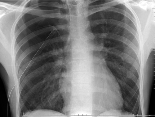
Looks perfect! But it’s a 2-D view and you don’t know where the tube is in the anterior-posterior axis. It turns out to be in the patient’s subcutaneous tissues of his back, near his scapula!

So what if this is a trauma activation patient and you are getting ready to send your patient for a chest CT shortly? Should you follow the usual dogma and still get a conventional chest x-ray prior to leaving the trauma bay?

The answer is no! Typically, your trauma activation patient should have rapid access to the CT scanner, so you won’t have to wait very long. And the additional 3-D information is very helpful in making sure the tube is placed exactly where you want it.

Bottom line: If you are planning on obtaining a chest CT anyway in your trauma patient, don’t bother with a conventional chest x-ray first to check chest tube position. But DON’T order a chest CT for this reason alone! Remember, the chest CT is only for detecting aortic injury in blunt trauma. It should not be used for diagnosing fractures, hemothorax, or pneumothorax. Or chest tube position!

Related posts:


Quiz: Is This A Good Chest Tube?

A blunt trauma activation patient presents with a pneumothorax seen on the initial chest x-ray, obtained in your trauma bay. You professionally insert a large chest tube, and all appears to go well. You shoot a followup chest x-ray and this is what you get:

What do you think of the tube position? Looks great, right?

But if you look carefully, you can see the lung outline in the middle of the right side of the chest. Big-time pneumothorax despite what looks like a perfectly placed tube. There are several possible explanations, and many of you sent me your guesses:

  • The tube is in the lung. This rarely happens to normal lungs. Sure, you can probably do it to an ARDS lung, but otherwise it’s not very likely.
  • The tube is in the fissure. This does happen on occasion, but not often. And many times it works anyway.
  • The tube is occluded or kinked. A PA or AP chest x-ray will show the kink, although bent tubes frequently work anyway. If a hemothorax is present, it is possible that a clot is plugging the tube. Clearing a plugged tube will be the subject of another post.
  • It’s not really a chest tube. Hopefully, this would have been detected when it was placed, but it isn’t always. The chest x-ray above looks great, right? Unfortunately, it’s a 2 dimensional representation of a 3-D object. Where is that tube in the z-axis?

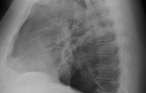
In this case the correct answer is the last one. This is one time when I would actually recommend a lateral chest x-ray. Have a look at the result. You can clearly see the tube snaking around into the soft tissues of the back.

Bottom line: Remember that a perfect x-ray doesn’t necessarily mean a perfect tube. Go through the various possibilities quickly, and make it work.

Related posts: