All posts by The Trauma Pro

Trauma Surgery Tip: How To See The Unseeable

Let me present a scenario and first see how you might solve this problem.

A young man presents with a gunshot to the abdomen in the right mid-back. He is hemodynamically stable, and you get a chest xray. It shows a small caliber slug in the right upper quadrant, but no hemo- or pneumothorax. He has peritoneal signs, so you whisk him off to the OR for a laparotomy.

As you prep the patient for the case, you can feel a small mass just above the right costal margin. You incise the area and produce a 22 caliber bullet. Of course, you follow the chain of evidence rules and pass it off for the police. As you explore the abdomen, it appears that there are no gross injuries. You are concerned, however, that there may be an injury to the diaphragm in proximity to the bullet.

So here’s the question: how can you visualize the diaphragm in this area? The bullet was located below the right nipple. But the diaphragm in this area is covered by the liver, and is parallel to the floor. You can’t seem to feel a hole with your fat finger. But short of putting your whole head in the wound, you just can’t get a good angle to see the area in question.

How would you do it? Please tweet or leave comments with your suggestions. I’ll provide the answer(s) tomorrow!

Liquid Plasma vs FFP: Impact On Your Massive Transfusion Protocol

In my last post, I discussed the growing number of choices for plasma replacement. Today I’ll look at some work that was done that tried to determine if any one of them is better than the others when used for the massive transfusion protocol (MTP).

As noted last time, fresh frozen plasma (frozen within 8 hours, FFP) and frozen plasma (frozen within 24 hours, FP) have a shelf life of 5 days once thawed. Liquid plasma (never frozen, LQP) is good for the 21 days after the original unit was donated, plus the same 5 days, for a total of 26 days.

LQP is not used at most US trauma centers. It is more commonly used in Europe, and a study there suggested that the use of thawed plasma increased short term mortality when compared to liquid plasma. To look at this phenomenon more closely, a group from UTHSC Houston and LSU measured hemostatic profiles on both types of plasma at varying times during their useful life.

All products were analyzed with thromboelastography (TEG) and thrombogram, and platelet count and microparticles, clotting factors, and natural coagulation inhibitors were measured. They chose 10 units of thawed FFP and 10 units of LQP, and assayed them every 5 days during their useful shelf life.

Here are the factoids:

  • Platelet counts were much higher in day 0 LQP (75K) vs day 0 thawed plasma (7.5K). Even at end of shelf life, the LQP was 1.5x higher than thawed (15K vs 10K).
  • Thrombogram showed that LQP had higher endogenous thrombin production until end of shelf life
  • TEG demonstrated that LQP had a higher capacity to clot that gradually declined over time. It became similar to thawed plasma at the end of its shelf life.
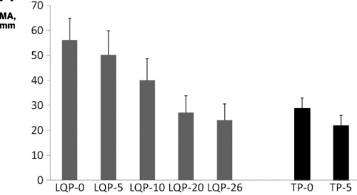
                         (TEG MA for liquid (LQP) and thawed (TP) plasma
  • Most clotting factors remained stable in LQP, with the exception of Factors V and VIII, which slowly declined

Bottom line: Liquid plasma sounds like good stuff, right? Although there are a few flaws in the collection aspect of this study, it gives good evidence that never frozen plasma has better coagulation properties when compared to thawed plasma. Will this translate into better survival when used in the MTP for trauma? One would think so, but you never really know until you try it. Our hospital blood bank infrastructure isn’t prepared to handle this product yet, for the most part. What we really need is a study that shows the survival advantage when using liquid plasma compared to thawed. But don’t hold your breath. It will take a large number of patients and some fancy statistical analysis to demonstrate this. I think we’ll have to look to our military colleagues to pull this one off!

Reference: Better hemostatic profiles of never-frozen liquid plasma compared with thawed fresh frozen plasma. J Trauma 74(1):84-91, 2013.

Liquid Plasma vs FFP: Definitions

I’ll spend the next few days discussing plasma. This is an important component of any trauma center’s massive transfusion protocol (MTP). Coagulopathy is the enemy of any seriously injured patient, and this product is used to attempt to fix that problem.

And now there are two flavors available: liquid plasma and fresh frozen plasma. But there is often confusion when discussing these products, especially when there are really three flavors! Let’s review what they are exactly, how they are similar, and how they differ.

Fresh frozen plasma (FFP)
This is plasma that is separated from donated whole blood. It is generally frozen within 8 hours, and is called FFP. However, in some cases it may not be frozen for a few more hours (not to exceed 24 hours total) and in that case, is called FP24 or FP. It is functionally identical to FFP. But note that the first “F” is missing. Since it has gone beyond the 8 hour mark, it is no longer considered “fresh.” To be useful in your MTP, it must be thawed, and this takes 20-40 minutes, depending on technique.

Thawed plasma
Take a frozen unit of FFP or FP, thaw, and keep it in the refrigerator. Readily available, right? However, the clock begins ticking until this unit expires after 5 days. Many hospital blood banks keep this product available for the massive transfusion protocol, especially if other hospital services are busy enough to use it if it is getting close to expiration. Waste is bad, and expensive!

Liquid plasma (never frozen)
This is prepared by taking the plasma that was separated from the donated blood and putting it in the refrigerator, not the freezer. It’s shelf life is that of the unit of whole blood it was taken from (21 days), plus another 5, for a total of 26 days. This product used to be a rarity, but is becoming more common because of its longer shelf life compared to thawed plasma.

Finally, a word on plasma compatibility. ABO compatibility is still a concern, but Rh is not. There are no red cells in the plasma to carry any of the antigens. However, plasma is loaded with A and/or B antibodies based on the donor’s blood type. So the compatibility chart is reversed compared to what you are accustomed to when giving red cells.

Remember, you are delivering antibodies with plasma and not antigens. So a Type A donor will have only Type B antibodies floating around in their plasma. This makes it incompatible with people with blood types B or AB.

Type O red cells are the universal donor type because the cells have no antigens on the surface. Since Type AB donors have both antigens on their red cells, they have no antibodies in their plasma. This makes AB plasma is the universal donor type. Weird, huh? Here’s a compatibility chart for plasma.

Next time, I’ll discuss the virtues of the various types of plasma when used for massive transfusion in trauma.

Vascular Trauma Resources At Pediatric Trauma Centers

There are two types of pediatric trauma centers: freestanding and combined. These adjectives refer to whether an adult trauma center is directly associated with the pediatric one. Over the years, I have come to appreciate that there may be substantial resource and experience differences between the two.

Trauma surgeons at freestanding centers are usually pediatric surgeons. They have managed trauma cases during their surgical residency and pediatric surgical fellowship, but usually have not taken a trauma fellowship. Their experience with complex trauma and advanced concepts like damage control surgery generally comes from their training and on the job experience. Surgeons at combined centers may be pediatric trained, or may be adult surgeons with pediatric experience. The adult surgeons are generally well-versed in advanced trauma concepts, and the pediatric surgeons can take advantage of the adult surgeons’ expertise in advanced trauma cases.

Freestanding pediatric centers may have fewer resources in some key areas, such as fellowship trained specialists in vascular surgery, GI endoscopy, and interventional radiology. A recent study accepted for publication from the University of Arkansas examines differences in surgeon practice patterns and resource availability at freestanding vs combined centers.

Two surveys were sent to 85 pediatric trauma centers around the US. Roughly half were Level I, and half were freestanding. One was sent to 414 pediatric surgeons at those centers inquiring about practice patterns, and the other was sent to the trauma medical directors of each center asking about their resources.

Here are the factoids:

  • 50 of the 85 trauma centers responded, as did 176 of the 414 surgeons. 48% of trauma medical directors responded. These are reasonable response rates for questionnaires.
  • Adult surgeons covered pediatric trauma at 6% of Level I centers, and 33% of Level II
  • During pediatric surgical fellowship, 56% participated in management of vascular trauma, 25% was managed by vascular surgeons, and 19% had no experience
  • At 23% of freestanding centers, vascular surgeons were not always available, and a vascular surgeon was not listed on the call schedule 38% of the time
  • 27% of freestanding facilities indicated that endovascular and stent capabilities were not available, and 18% did not have interventional radiologists (IR) available within 30 minutes
  • All combined centers had vascular and endovascular capabilities, and IR was available within 30 minutes 92% of the time

Bottom line: This is an intriguing paper that looks at a few of the disparities between freestanding and combined pediatric trauma centers. Obviously, it is hampered by the survey format, but does provide some interesting information. The focus was on vascular resources, and shows several of the major differences between the two types of centers.

Fortunately, vascular trauma is relatively rare in the pediatric age group. But it is possible that a child presenting to a freestanding pediatric trauma center may be managed by a pediatric surgeon with little vascular experience, and assistance from a fellowship trained vascular surgeon and/or interventional radiologist may be unavailable.

This paper provides important information regarding resource disparities in pediatric trauma care. Ideally, this should be reviewed and remedied as the Resources for Optimal Care of the Injured Patient (Orange Book) evolves over the coming years.

Reference: Pediatric Vascular Trauma Practice Patterns and Resource Availability: A Survey of ACS-Designated Pediatric Trauma Centers. J Trauma, accepted for publication Jan 12, 2018.

The Final X-Ray In Damage Control Surgery

Damage control surgery for trauma is over 20 years old, yet we continue to find ways to refine it and make it better. Many lives have been saved over the years, but we’ve also discovered new questions. How soon should the patient go back for definitive closure? What is the optimal closure technique? What if it still won’t close?

One other troublesome issue surfaced as well. We discovered that it is entirely possible to leave things behind. Retained foreign bodies are the bane of any surgeon, and many, many systems are in place to avoid them. However, many of these processes are not possible in emergent trauma surgery. Preop instrument counts cannot be done. Handfuls of uncounted sponges may be packed into the wound.

I was only able to find one paper describing how often things are left behind in damage control surgery (see reference below), and it was uncommon in this single center study (3 cases out of about 2500 patients). However, it can be catastrophic, causing sepsis, physical damage to adjacent organs, and the risk of performing an additional operation in a sick trauma patient.

So what can we do to reduce the risk, hopefully to zero? Here are my  recommendations:

  • For busy centers that do frequent laparotomy or thoracotomy for trauma and have packs open and ready, pre-count all instruments and document it
  • Pre-count a set number of laparotomy pads into the packs
  • Use only items that are radiopaque or have a marker embedded in them. This includes surgical towels, too!
  • Implement a damage control closure x-ray policy. When the patient returns to OR and the surgeons are ready to begin the final closure, obtain an x-ray of the entire area that was operated upon. This must be performed and read before the closure is complete so that any identified retained objects can be removed.

Tomorrow, a sample damage control closure x-ray.

Related post:

Reference: Retained foreign bodies after emergent trauma surgery: incidence after 2526 cavitary explorations. Am Surg 73(10):1031-1034, 2007.