All posts by The Trauma Pro

Ever Wonder Where The Golden Hour Came From?

Everywhere you turn in the trauma and EMS world, you run into the concept of the “golden hour.” Basically, it refers to the idea that it’s important to get an injured patient to definitive care promptly, or mortality begins to rise. It has been used to justify a lot of what we do in trauma care and trauma systems. But where did this come from? And is it true?

The BTLS course attributes the term to R Adams Cowley from the ShockTrauma Center in Baltimore. Unfortunately, no references are given. A biography of Cowley entitled Shock-Trauma names him the author of the term, basing it on dog research. No references were given.

A review of Cowley’s research reveals a few tidbits. A case series of patients implies that speed is good, but does not analyze time to definitive care. It does reference older work by other authors, but once again, no relationship between timing and outcome is evaluated.

A textbook edited by Cowley contains a reference to an article about “Cowley’s golden hour.” This article contains a statement that “patients are assumed to be dying and much of the golden hour has passed.” It goes on to state that the first 60 minutes after injury determines the patient’s mortality. It, in turn, refers to another of his earlier articles. This one states that “the first hour after injury will largely determine a critically injured person’s chance for survival.” No data or reference is given.

Bottom line: The concept of the “golden hour” has taken on a life of its own. Yes, it’s a good idea. And yes, there is some actual data to support it, although the quality is somewhat lacking. But this does point out the need to question everything, even some of our most deeply held beliefs. They are not always what they seem to be.

Reference: The Golden Hour: scientific fact or medical urban legend? Acad Emerg Med 8(7):758-760, 2001.

Complications Of Iliac Artery Embolization

The main cause of mortality in patients with severe pelvic fractures is major hemorrhage. Over the years, trauma professionals have developed and tested a number of maneuvers to reduce mortality in these patients. These include wrapping or fixing the pelvis, embolization, and more recently, pre-peritoneal packing and REBOA.

Pelvic wrap/fixation and embolization have been around for a long, long time. For both, it’s been long enough so that we should have a fairly decent appreciation of the complications. For pelvic binders, they principally involve the skin. But aside for the potential access site complications (bleeding, pseudoaneurysm), angiography has been thought to be relatively benign.

But as with any medical procedure, especially invasive ones, there are risks. A paper published five years ago retrospectively reviewed the 13 year experience with pelvic angiography at UC Davis. Study patients were matched with controls who underwent angiography for pelvic fracture but not embolization. Short-term (within 30 days) and long-term complications were assessed while in hospital and by telephone survey. Mean followup time was 18 months.

Here are the factoids:

  • There were no differences in complications attributable to embolization within 30 days of the procedure
  • There were 5 cases of short-term skin sloughing or necrosis in 55 patients, and 4 of 5 occurred in patients with nonselective embolization. However, this was not a statistically significant complication.
  • Long-term complications such as buttock claudication or skin ulceration, pain, and impotence were not significantly different in embolized vs non-embolized patients
  • There was a significantly increased incidence of buttock, perineal, or thigh paresthesias in the long-term

Bottom line: Angiography with embolization is a very valuable tool in the management of complication pelvic fractures. Remember that a number of complications have been described:

  • Skin sloughing or necrosis
  • Buttock claudication, pain, paresthesias
  • Skin ulceration
  • Impotence

Other than an increase in paresthesias in the long-term, there did not appear that there was any difference in patients undergoing angiography with and without embolization. Although the numbers were small (100 patients total), this is the best study we have to date. Just keep in mind that complications are possible, and question your patients about them when they present for their followup visits.

Reference: Evaluation of Short-term and Long-term Complications after Emergent Internal Iliac Artery Embolization in Patients with Pelvic Trauma. J Vascular Interventional Rad 19(6):840-847, 2008.

Part 2: Metal Splints – Can You CT Scan Through Them?

In my last post, I debunked the myth that using a pre-formed aluminum splint significantly degrades the quality of standard x-rays. But what about a study that provides much more detail, such as CT scan?

CT scan techs have told me that there would be too much artifact using any kind of metal splint. And typically, when imaging an extremity with CT, we are looking at vascular runoff. The vessels are small, and high image quality is extremely important. If the images are bad, then we risk having to give the patient another dose of both radiation and contrast.

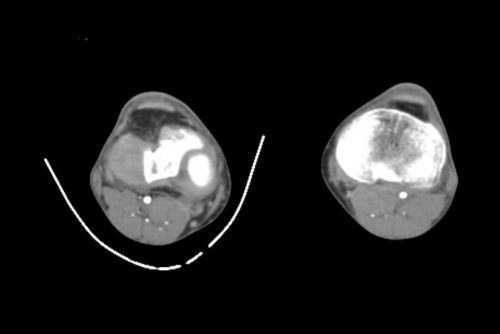
As you know, my mantra is question everything! So i scouted around and found some images to share using one of these splints. Look closely for the intimal flap in the image below:

Can’t see it? That’s because it isn’t there! But you certainly could if it were!

Bottom line: A perforated aluminum splint causes absolutely no artifact or image degradation. Do not cause additional injury by removing it prior to imaging, either CT or conventional x-ray. Although your friendly techs, radiologists, and orthopedic surgeons may moan, it won’t hurt their ability to make decisions on the images.

Part 1: Metal Splints – Can You X-ray Through Them?

Splinting is an important part of the trauma resuscitation process. No patient should leave your trauma resuscitation room without splinting of all major fractures. It reduces pain, bleeding, and soft tissue injury, and can keep a closed fracture from becoming an open one.

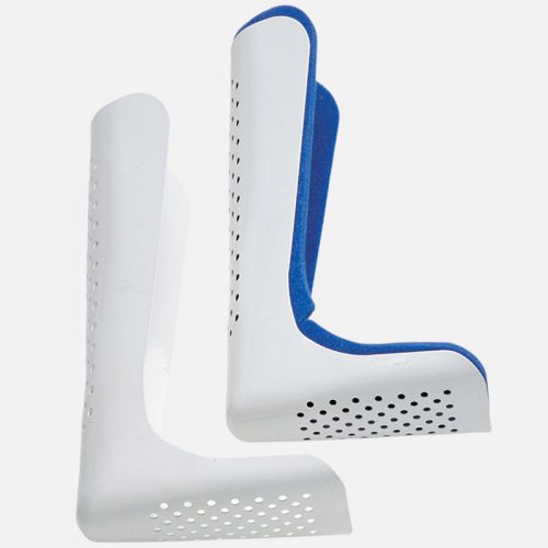
But what about imaging? Can’t the splint degrade x-rays and hamper interpretation of the fracture images? Especially those pre-formed aluminum ones with the holes in them? It’s metal, after all.

Some of my orthopedic colleagues insist that the splint be removed in the x-ray department before obtaining images. And who ends up doing it? The poor radiographic tech, who has no training in fracture immobilization and can’t provide additional pain control on their own.

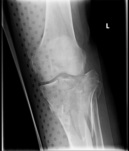
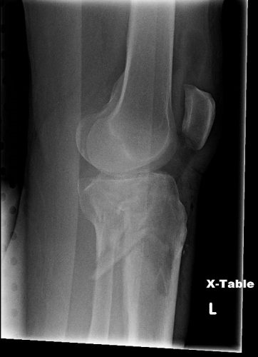
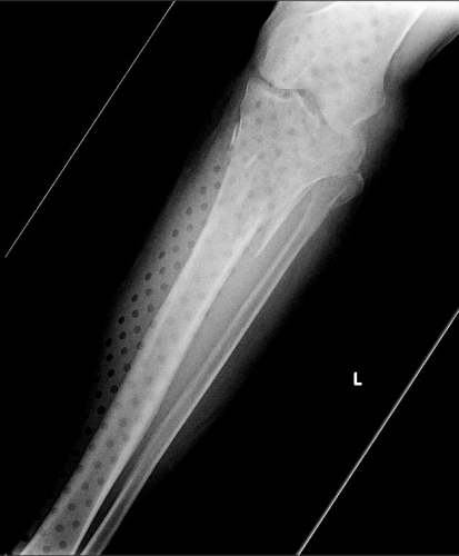
But does it really make a difference? Judge for yourself. Here are some knee images with one of these splints on:

Amazingly, this thin aluminum shows up only faintly. There is minimal impact on interpretation of the tibial plateau. And on the lateral view, the splint is well posterior to bones.

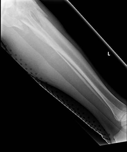
On the tib-fib above, the holes are a little distracting on the AP view, but still allow for good images to be obtained.

Bottom line: In general, splints should not be removed during the imaging process for acute trauma. For most fractures, the images obtained are more than adequate to define the injury and formulate a treatment plan. If the fracture pattern is complex, it may be helpful to temporarily remove it, but this should only be done by a physician who can ensure the fracture site is handled properly. In some cases, CT scan may be more helpful and does not require splint removal. And in all cases, the splint should also be replaced immediately at the end of the study.

In my next post, I’ll look at the use of CT scans when this type of splint is in use.