All posts by The Trauma Pro

Where Did The Hypodermic Needle Gauge System Come From?

Yesterday, I wrote about one of the weirder measurement systems in medicine, the French (or Charrière) system for catheter sizes. Today, I’ll deal with another arcane system, the gauge system for hypodermic needles.

I’ve always wondered how this worked. Lower numbers somehow indicate bigger needles. And to make it more confusing, there are two conflicting gauge systems, the Birmingham Wire Gauge (aka the Stubs Iron Wire Gauge) and the Stubs Steel Wire Gauge. Confusing, right? And these two are actually a little different.

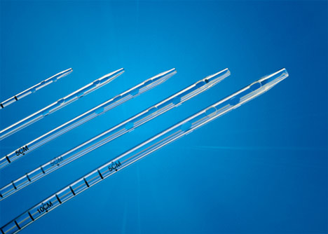
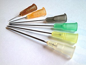
Hypodermic needle sizes are based on the Birmingham gauge system (G).  The Gauge indicates the outer diameter (OD) of the wire used to make the needle, but is inversely related to it. And to top it off, there is no real mathematical formula that relates gauge to the OD. If you are that interested, you have to consult a printed table.

Needle gauge ranges from 7G (4.57mm OD) to 34G (0.18mm OD). There is also little correlation of the inner diameter to gauge, as the wall thickness decreases as the OD of the needle decreases. As an interesting tidbit, there is an International Standards Organization (ISO) standard for single-use needles that determines the color coding of the hub. I always wondered who thought those up!

Okay, so needle gauge is based on the size of the wire used to make them. What about IV catheters? Just to make it more confusing,  IV catheter size indicates the size of the needle used to insert them. So an 18G IV catheter has an inner diameter that is almost identical to the OD of its needle. The outside diameter of the catheter will be a tiny bit bigger. Got it?!

So there you have it. Amaze your friends with your arcane knowledge about tubes and needles and catheters!

Where Did The French Tube Size System Come From?

Medicine sure has some weird measurement systems. Besides the more standardized units like microliters, milligrams, and International Units, we’ve got some odd stuff like French (tubes) and gauge (needles). When dealing with tubes and catheters, the size is usually specified in French units.

Where did the French system come from? It was introduced by a Swiss-born gentleman named Joseph-Frédéric-Benoît Charrière. He moved to Paris and was apprenticed to a knife maker. At the age of 17, he founded a  company that manufactured surgical instruments. His company developed and improved a number of surgical instruments, including hypodermic needles and various catheters.

Charrière introduced the system for describing catheters based on their outer diameter (OD).  It was actually named after him, and in France one will occasionally see catheters described in Ch units. Unfortunately, we Americans had a hard time pronouncing his name, and changed it to the French system (Fr).

So what’s the translation? The Ch or Fr number is the outer diameter of a catheter in millimeters multiplied by 3. It is not the outer circumference in millimeters, and the use of pi is not involved. So a big chest tube (36 Fr) has an OD of 12 mm, and a bigger chest tube (40 Fr) has an OD of 13.33 mm.

Tomorrow: Where did the needle gauge size come from?

Chest Tubes: Size Doesn’t Matter – Part 2

A few days ago, I wrote about a paper that seemed to suggest that using a smaller chest tube (28-32 Fr) vs larger ones (36-40 Fr). The results suggested that their function was very similar. I emphasized that I thought the result was intriguing, because I’m of the opinion that bigger is better for getting clotted blood out. However, I am amenable to changing my mind based on newer, better data.

But I did caution readers that I would like to see more data. One study should never change your practice! Then I see a lot of chatter on Twitter about another study from 2016 that looks at even smaller tubes, with people saying they will now switch to pigtail catheters (12 Fr)!!

First, not a logical progression of thinking there. And second, let’s take an actual look at the paper. It’s from an emergency medicine group in Fukui, Japan, which retrospectively reviewed their 7 year experience with using a small (20-22 Fr) vs large (28 Fr) tubes. They identified a total of 124 chest tube insertions to compare, 68 small and 56 large.

Now let’s look at the factoids:

  • Demographics, mechanism, and ISS were the same between groups
  • Duration of insertion and initial drainage were also the same between groups
  • Complication rates were similar, with 1 empyema and 2 retained hemothoraces in each group
  • Additional tubes were place in 2 patients with small tubes vs 4 with large tubes
  • Thoracotomy was performed in 2 patients with small tubes vs 1 with a large tube

Based on all of this, the authors concluded that there was no difference in drainage efficacy, complications, or need for additional invasive procedures.

Wait a minute!! Again, if you only read the abstract, you might be led to start using ever smaller chest tubes. But read the entire paper! There are many problems with this paper, including:

  • It’s a very small, retrospective review. This automatically means that the statistical power is suspect.
  • Why did they only document 124 insertions over 7 years?? That’s about one every 3 weeks! Either a lot of data are missing or they are not very busy. But Fukui Prefectural Hospital has over 1000 beds! So it’s the former, not the latter.
  • The retrospective nature means it is not possible to determine why a particular tube size was chosen. Roll of the dice? This fact alone introduces a huge potential for selection bias. Was a smaller tube selected because the hemothorax looked smaller? Probably! The fact that 4 patients with larger tubes had another one placed suggests that they were being used for larger collections. And patients with higher ISS tended to get bigger tubes.

Bottom line: Don’t change your practice based on this paper. And certainly don’t choose to use even smaller pigtails. And of course, always critically read any paper that you like to make sure you are not cherry picking the ones you choose to believe. IMHO, it’s still best to use big (36 Fr) or bigger (40 Fr).

Reference: Small tube thoracostomy (20-22 Fr) in emergent management of chest trauma. Injury 48:1884-1887, 2016.

Trauma Mythbusters: NSAIDs And Fracture Healing

Trauma hurts like hell. Over the years, we’ve developed quite a few ways of combating this pain. A number of drug classes have been developed to reduce it. One of the more common non-narcotic drug classes are the NSAIDs. As I’ve mentioned before, every drug has dozens of effects. Drug companies market a particular medication based on one of the predominant effects. All the others are considered side effects.

NSAIDs are not unique; they have lots of side effects as well. In 2003, several papers brought to light possible interactions between these drugs and fracture healing. Specifically, there were questions about these drugs interfering with the healing process and of increasing the number of delayed unions or nonunions. But once again, how convincing were these papers, really?

It would seem to make sense that NSAIDs could interfere with bone healing. This  process relies heavily on the regulation of osteoblast and osteoclast function, which itself is regulated by prostaglandins. Since prostaglandins are synthesized by the COX enzymes, COX inhibitors like the NSAIDs should have the potential to impair this process. Indeed, animal studies in rats and rabbits seem to bear this out.

But as we have seen before, good animal studies don’t always translate well to human experience. Although a study from 2005 suggested that NSAID administration in older patients within 90 days of injury had a higher incidence of fracture nonunion, the study design was not a very good one. It is equally likely that patients who required these drugs in this age group may have been at higher risk for nonunion in the first place.

In fact, there are no large, prospective randomized studies that have explored the effect of short-term or long-term NSAID administration on fracture repair. But there have been several smaller studies that showed absolutely no effect on nonunion with short-term administration of this drug class. Yet the dogma that leads us to avoid giving these drugs persists.

A recent analysis looked at the quality of the published research, both for and against NSAID usage in fracture patients. They used the Coleman Methodology Score, which evaluates study size and type, mean followup, detailed description of treatment, subject selection, outcomes, and outcome assessment. The maximum score was 100. 

Here are the factoids:

  • There were 4x as many total subjects in the “NSAIDS are okay” papers than in the “avoid NSAIDS” papers
  • The quality of the “NSAIDS are okay” papers were significantly higher than “avoid NSAIDS” group (59 vs 40)
  • Interestingly, the “avoid NSAIDS” papers are cited twice as often
  • All of the reviews ended with my pet peeve catch phrase “further (good) research is needed”

Bottom line: Once again, the animal data is clear but the human data is not. Although there are theoretical concerns about their use, there is not enough solid risk:benefit information to abandon short-term NSAID use in patients who really need them. NSAIDs can and should be prescribed in patients with short-term needs and simple fractures.

References:

  1. Effects of nonsteroidal anti-inflammatory drugs on bone formation and soft-tissue healing. J AM Acad Orthop Surg 12:139-43, 2004.
  2. Effect of COX-2 on fracture-healing in the rat femur. J Bone Joint Surg Am 86:116-123, 2004.
  3. Effects of perioperative anti-inflammatory and immunomodulating therapy on surgical wound healing. Pharmacotherapy 25:1566-1591, 2005.
  4. Pharmacological agents and impairment of fracture healing: what is the evidence? Injury 39:384-394, 2008.
  5. High dose nonsteroidal anti-inflammatory drugs compromise spinal fusion. Can J Anaesth 52:506-512, 2005.
  6. Nonsteroidal Anti-Inflammatory Drugs and Bone-Healing: A Systematic Review of Research Quality. JBJS Rev 4(3), 2016.
What Does A Retained Surgical Sponge Look Like?

Surgeons and surgical residents rarely see these. And because it’s so uncommon, they frequently don’t recognize the telltale findings on radiographic studies. The TSA runs into the same problem in screening passengers for weapons and other hazards at airports. But it’s the bane of any surgeon’s existence. And it’s a major reason why OR personnel take such great pains to account for everything in the room. It is a catastrophe, and always a preventable one, when some piece of equipment goes missing and ends up left inside a patient.

A number of methods have been developed to try to eliminate this problem. They include careful counts, having someone record anytime anything is placed inside, x-rays, and most recently, RFID tags.

After counting, x-ray is the most common way to try to find missing objects. One would think that these foreign bodies would be easy to see. Metallic instruments are rather easy to spot. But many trauma professionals, even those who work in the OR, have never seen what a positive image of a sponge actually looks like. So here they are. You should never miss one on an xray now.

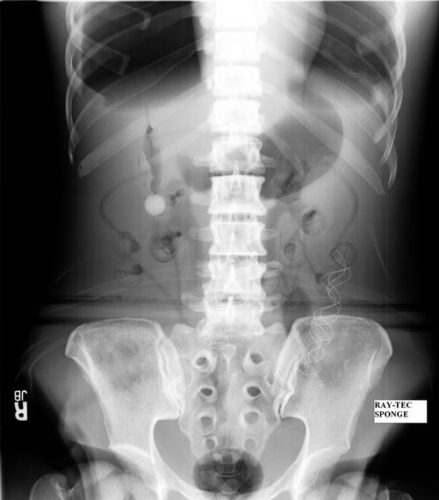
Surgeons typically use two types of sponges in the OR: Ray-Tec sponges and standard lap pads. Ray-Tecs look like a 4×8 piece of gauze with a mysterious blue string woven throughout it. The string is the only part that shows up on x-ray, and it is very thin and somewhat hard to see. Here are some Ray-Tec sponges outside the body:

And here’s one that was left inside. Note the little squiggle in the left lower quadrant and how easy it is to overlook.

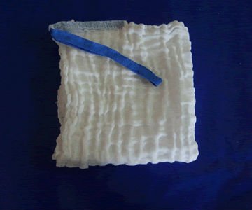
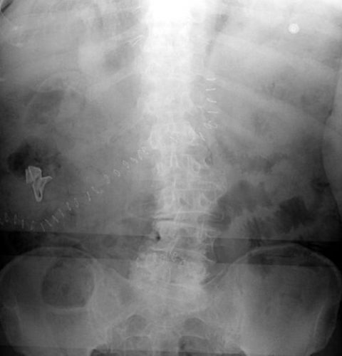
On the other hand, a laparotomy pad is a 4×4 folded cloth pad that unfolds into a larger pad. It has a blue radiopaque tag sewn in the corner, extending along one edge of the pad. Here’s what they look like outside the body:

And here’s one inside a patient. Note the irregular object in the right upper quadrant. Many times the tag is scrunched up and doesn’t look like one.

Bottom line: It’s important for anyone who works in the OR on any body part to be familiar with the appearance of these tags on x-rays. Since it’s generally impossible to get accurate counts before or after a trauma procedure, always image the involved body cavity looking for these telltale signs before closing the patient.

Note: These images  were taken from the internet. Patients were not treated at Regions Hospital.