All posts by The Trauma Pro

Solid Organ Injury Practice Guideline Updated

Regions Hospital developed a clinical practice guideline for solid organ management in 2002-2003. It has been revised a few times over the years, as any good guideline should with the availability of new data.

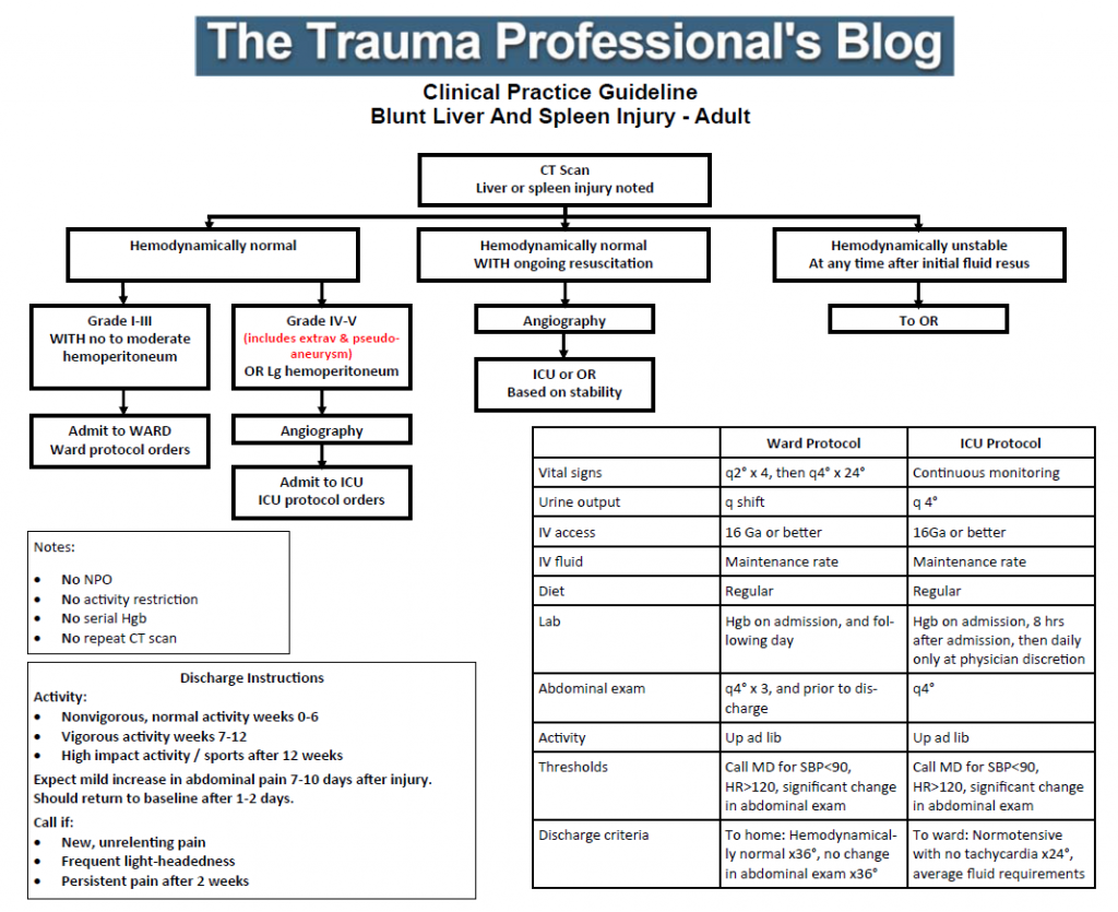
I’ve just put the finishing touches on the latest revision as a result of the updated organ scaling rules published by the American Association for the Surgery of Trauma. I reviewed the new scales for both liver and spleen earlier this year (links below). In the previous iteration of the scaling system, the importance of contrast pooling (pseudoaneurysm) or extravasation beyond the organ was not well defined. 

The new guideline explicitly includes these injuries in the high grade group, which for us is grade IV or V. Technically, pseudoaneurysm of the liver is only grade III, but in my opinion demands angiographic investigation and embolism. Thus the inclusion in the high grade / angiography arm of our guideline.

For those of you who have not seen this guideline before, there are several important directives that are listed on the left side of the page:

  • Patients are NOT made NPO
  • They do NOT have activity restrictions (such as bed rest)
  • Serial hemoglins are NOT drawn
  • An abdominal CT scan is NOT repeated

These changes were made in 2015 based on our clinical experience that properly selected patients almost never failAnd they still don’t, so why starve, restrain, poke, and re-irradiate them?

Additionally, we included explicit impact activity restrictions for post-discharge so that patients would get the same message from all members of our team.

Click the image below to download the guideline and have a look. I’m interested in your comments!

Related posts:

Tension Pneumothorax From Inside The Chest: The Video

This video shows what it looks like from the inside when a needle thoracostomy is inserted into the chest. Note that it takes about a minute for the lung to expand, so be patient when you insert the needle. You can also get an idea of why the needle is only a temporary measure as the inflating lung begins to kink the catheter.

Don’t ask why there was a thoracoscope in the chest with a tension pneumo in the first place, though!

YouTube player

IV Contrast and Trauma – Revisited

We use CT scanning in trauma care so much that we tend to take it (and its safety) for granted. I’ve written quite a bit about thoughtful use of radiographic studies to achieve a reasonable patient exposure to xrays. But another thing to think about is the use of IV contrast.

IV contrast is a hyperosmolar solution that contains some substance (usually an iodine compound) that is radiopaque to some degree. It has been shown to have a significant impact on short-term kidney function and in some cases can cause renal failure.

Here are some facts you need to know:

  • Contrast nephrotoxicity is defined as a 25% increase in serum creatinine, usually within the first 3 days after administration
  • There is usually normal urine output and minimal to no proteinuria
  • In most cases, renal function returns to normal after 3-4 days
  • Nephrotoxicity almost never occurs in people with normal baseline kidney function
  • Large or repeated doses given within 72 hours greatly increase risk for toxicity
  • Old age and pre-existing diabetic renal impairment also greatly increase risk

If you must give contrast to a patient who is at risk, make sure they are volume expanded (tough in trauma patients), or consider giving acetylcysteine or using isosmolar contrast (controversial, may still cause toxicity).

Bottom line: If you are considering contrast CT, try to get a history to see if the patient is at risk for nephrotoxicity. Also consider all of the studies that will be needed and try to consolidate your contrast dosing. For example, you can get CT chest/abdomen/pelvis and CT angio of the neck with one contrast bolus. Consider low dose contrast injection if the patient needs formal angiographic studies in the IR suite. And finally, consider what changes will be made if the study is positive. For example, if a CT angio of the neck for blunt carotid/vertebral injury is being considered, the intervention for a positive result is usually just aspirin. Since this is a very benign medication, why not forgo the scan and just start aspirin if there is a significant risk of kidney injury from the contrast. Always think about the global needs of your patient and plan accordingly (and safely).

Reference: Contrast media and the kidney. British J Radiol 76:513-518, 2003.

Button Batteries: Part 2 – Getting Them Out

In my last post, I detailed how to suspect and image a button battery ingestion. In this one, I’ll describe how to extract them, and how quickly it’s necessary.

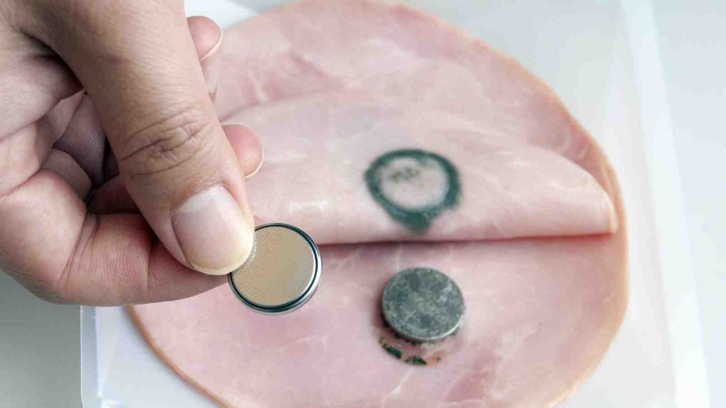
When batteries come to rest and are surrounded by moist mucosal tissue, a current arc is generated around the two sides of the button. This releases heat, which coagulates the surrounding tissue. Depending on the location, closeness of contact, and the duration, these burn injuries may extend into underlying tissue. This is of particular significance in the esophagus, which is in close proximity to the thoracic aorta.

Here’s a simple demonstration you can do at home with some lunch meat:

Here are guidelines for what to do when you encounter pediatric patients who have ingested a button battery:

  • If the child is experiencing bleeding from the upper GI tract, activate your trauma team. The child may have an aorto-esophageal fistula. If there is no active bleeding, obtain a chest x-ray to assess the battery’s position. If there is active bleeding, proceed to the OR (preferably a hybrid room if you have one) and use fluoro to locate the battery. If bleeding persists, call appropriate pediatric surgical specialists (surgery, CV surgery, GI), activate your massive transfusion protocol, and consider tamponade with a Blakemore tube (remember those?) or a urinary catheter if you don’t have one.
  • No bleeding from the upper GI tract? If the battery is large (>20mm) and/or the child is small (<5 years), and is lodged in the esophagus, proceed immediately to OR and remove endoscopically.
  • Batteries in the stomach are of less concern. They will generally pass if <20mm. A repeat x-ray after 48 hours should be obtained for larger batteries. If still in the stomach, they should be removed endoscopically. Smaller batteries will usually pass, and should be re-imaged after two weeks to confirm this.

References:

  • Button battery and magnet ingestions in the pediatric patient.  Curr Opin Pediatrics 30:653-659, 2018.
  • Management of ingested foreign bodies in children: a clinical report of the NASPGHAN Endoscopy Committee. J Pediatric Gastroenterol Nutr 60:562-574, 2015.