Tag Archives: what the heck?

What The Heck? The Answer

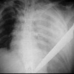
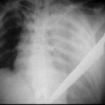
Obviously, there’s a big knife in this image, and it appears to coincide with our patient. Remember, you can’t tell the relative positions of objects in the z axis in a two dimensional radiograph. However, you know that it is not on top of or under this patient, because there is an obvious hemothorax.

First, this patient should have been a trauma activation, since there is a penetrating injury of the chest. The first decision point is based on vital signs: is the patient hemodynamically normal? If not, do the essentials quickly (quick exam, lines, blood draw for blood bank) and move to the OR. The quick exam is important, especially of the back, since you may not see it for the next few hours and additional injuries there could be trouble.

If the patient is stable, then it’s time to do the essentials and figure out what you are going to need to do in the next hour. A chest xray can be very helpful. However, given the size of the knife and the fact that it appears to be buried to the hilt allows you to calculate the trajectory in your mind. The only question is if it went in far enough to enter the abdomen.

The xray shows a moderate hemothorax and a tip that extends well into some part of the abdomen, no matter how you look at it. It has also come alarmingly close to the heart. If the knife had not penetrated as deeply and was obviously only in the chest, then removal of the knife with insertion of a chest tube would be appropriate. 

However, in the case of this xray, the patient still needs to go to the OR. The chest and abdomen need to be prepped and the abdomen opened first. Once the knife is located, the next steps can be planned. If there is concern for the heart, the pericardial surface of the diaphragm can be opened to look for blood. If found, extension of the laparotomy incision into a sternotomy is appropriate for cardiac repair. If not, the tip of the knife should be visualized and the weapon slowly removed under direct vision. Any bleeding structures can be dealt with first as it goes, followed by any injuries to a hollow viscus.

What The Heck? Final Answer

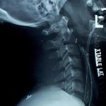
This one was a bit tricky. I chose it because it looks like there is an extra tube in the neck. You can see two stripes traveling from the mouth down the neck. The one closest to the cervical spine is in the esophagus, an orogastric tube. The other one passes anterior to it, in the trachea, so it is the orotracheal tube. But what about the tube shaped density that is located in the posterior pharynx that looks like it is angled forward toward the trachea? Did someone lose something?

If you think about it though, you should conclude it’s something weird. There is no radiopaque stripe on it, which rules out most common tubes. The only thing of that size and shape that comes to mind is a nasopharyngeal airway tube. However, these have a flange on the nasal end, so it couldn’t just pass inwards through the nose. And who in their right mind would put it in the mouth to be swallowed? Plus, the orientation of it is unusual, heading forward toward the trachea.

You have to look at the rest of the clues on the radiograph. It’s easy to get suckered if you just focus on the obvious. What are those objects located between the two tube stripes in front of C6? Surgical clips. What are those O-shaped objects at the angle of the mandible that disappear behind the XTABLE LAT marker? Surgical skin closure staples.

So this is a postoperative patient. If you follow the object, it actually moves toward the skin, and beyond! This patient was stabbed in the neck and underwent a surgical exploration with control of bleeding. A surgical drain was placed due to concern for leakage from the pharynx or salivary glands. The drain actually leaves the side of the neck, just anterior to the sternocleidomastoid muscle.

Remember to look at everything on a radiograph, especially if you don’t have the clinical story behind it. The eye normally focuses on the obvious, leading the viewer to make assumptions based on their expectations. This can easily get you in trouble, so beware! And don’t forget that you are looking at a 2-D image, so there is no way to tell where any object is in the third dimension. It may be in the front, the back, or under the patient in their clothing!