Tag Archives: what the heck?

What The Heck? The Answer!

Yesterday I posted an image of an unusual chest CT. The patient had been involved in a motorcycle crash weeks ago, and presented with new onset chest pain and weakness.

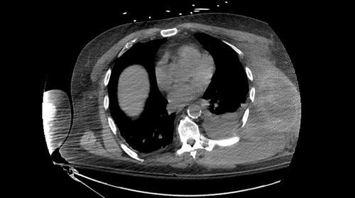
Exam of the chest showed a hint of diffuse swelling on the left side and moderate tenderness. Chest x-ray suggested a mild effusion on the left. I showed one slide of the CT yesterday, which showed a large amount of complex material in the chest wall. This is most likely a mixture of blood and clot. 

Here is another slice of the CT that is more revealing:

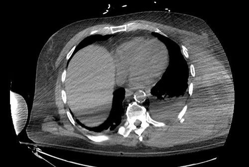
Now you can see that there are multiple rib fractures present. While comparing the original and the recent scan, it is apparent that the fractures are more displaced on the recent one. Upon closer questioning the patient admits that he did fall down the day before the new pain and swelling occurred.

And by the way, he forgot to mention the fact that he had developed deep venous thrombosis and was taking warfarin! And also by the way, his blood pressures were becoming a bit soft.

I would consider this life-threatening bleeding! Crystalloid and blood resuscitate immediately. Reverse the anticoaguation quickly, using prothrombin complex concentrate (PCC, preferably 4-factor). Then send him to interventional radiology to see if there are any active bleeders that can be embolized. Finally, it’s off to the ICU to finish up the resuscitation and restore him to normal!

Related posts:

What The Heck? Chest CT

Here’s another one for you to figure out. Of course, I’ll give you minimal information and see what you can do with it. 

A middle-aged male presents to your ED from home with left chest pain and weakness. There is a history of a motorcycle crash 6 weeks ago. Ultimately, a chest CT was obtained and here is a representative section:

Here are my questions:

  • What’s going on?
  • Why?
  • What should you do about it?

Leave a comment below or tweet your guesses. Answers tomorrow!

What The Heck? Some Hints!

Yesterday, I presented the case of a young man with abdominal pain a few weeks after a splenectomy for trauma. One slice of the CT scan was presented, which showed pneumatosis in the wall of the cecum.

There have been some great comments from readers with some interesting reasoning, especially from StillChucklesandNP. Here’s another piece of the puzzle, yet another slice from the CT scan:

  • What’s this?
  • Any other important information?
  • What’s the diagnosis and why?
  • How do you treat it?

Looking forward to more tweets and comments! Answers tomorrow.

What The Heck?

Here’s an interesting one for you to solve!

A 20ish year old male was involved in a motor vehicle crash, sustaining a Grade IV spleen injury. He fails nonoperative management early in his hospital course, undergoing a splenectomy 6 hours later.

He has an uneventful recovery and is ready for discharge after 5 days. His platelet count has plateaued at 600K. He presents to your ED 2 weeks later complaining of abdominal pain. On exam, he is diffusely but mildly tender. His subjective complaints appear to be a bit out of proportion to his exam.

Here is one slice from his CT scan. I’ve put a nice fat arrow on it to help out. But it won’t.

image

  • What does the scan show?
  • Why is it there?
  • What other key piece of information do you want to know?
  • Any other studies?
  • Then what?

Some hints tomorrow! Tweet your answers or leave comments below! Let’s see if anyone can figure this one out!

What The Heck? Bicycle Trauma Answer

So a young male jammed a handlebar into his abdomen, and a CT image demonstrating his problem was shown. But what did it actually show?

By now, you probably realize that clinical information is key. On exam, he had an obvious bulge in his left lower quadrant, more obvious with straining. Looking at the CT (now with a nice arrow), there is a problem over the left side of the abdomen. 

image

This child has so little fat, that it’s difficult to see the problem. If you track the thin layer of fat across the abdomen to the right side of the image, you’ll see that it disappears over the bowel gas. This represents a complete tear through all fascial layers, not just a Spigelian hernia as some readers guessed.

Management consisted of primary repair of the defect. An uneventful recovery can be expected. Unless more bicycle tricks are anticipated.

Reference: Traumatic handlebar hernia: a rare abdominal wall hernia. J Ped Surg 39(10):e20-e22, 2004.