Tag Archives: technology

New Technology: Help Brain Injured Patients To Talk

It is can be extremely difficult to communicate with some brain injured patients. Many have global damage that precludes the processing necessary to formulate thoughts. However, some may be able to think but can’t effectively make themselves understood. Patients with the “locked in” syndrome are a perfect example.

A company called NeuroVigil has developed technology and data analysis techniques for extracting a wealth of information from a single-channel EEG. The iBrain system uses two sensors that do not require being stuck to the head with adhesive. A simple elastic band can hold them in place. 

Last year, the company fitted the device on Stephen Hawking to begin testing and training the system to assist with his communication efforts. Currently, Hawking uses an IR sensor that detects twitches in his cheek. These are painstakingly translated into letters and then words that are spoken by a computer. The iBrain system is being trained to recognize words via his EEG patterns and should speed up his communication with the outside world.

If this technology pans out, it may be used to communicate with moderate to severely injured TBI patients who have expressive language problems. It could also be used to test for and communicate with patients who are “locked in.”

The video was recorded at TEDMED 2009. Much of the key information is presented beginning at 10:10 into the video.

I have no financial interest in NeuroVigil

New Technology: The End Of Handwashing?

All healthcare professionals are notoriously bad about washing their hands, especially doctors. A variety of things have been developed to help us keep our hands clean, including simple soap and water, barriers like gloves, and various gels and foams (which I swear I can taste in my mouth 10 minutes later, even though I’m pretty sure I’m not putting my fingers there).

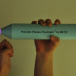
A recently published paper from China is shining new light on this topic (get it?). Researchers developed a hand-held, battery-powered plasma flashlight that gets rid of bacteria on skin in a flash. It costs less than $100 to produce and runs on a 12V battery. 

This device was found to inactivate all bacteria in a 17-layer biofilm containing a very hardy organism, enterococcus faecalis. It does not produce UV radiation, and the exact mechanism for the bacterocidal effect is unclear. There was no adverse effect on skin.

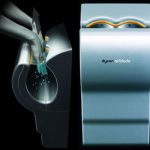
The main drawbacks to this device are that it only produces a small area of plasma, and it takes 5 minutes to kill all the bacteria. But take this to the next logical step. Many of you are familiar with the Dyson Airblade hand dryers found at many airports. Suppose you could produce a more intense plasma field using a more robust power supply (power line or ambulance power system) in a device that you could just pass your hands through to disinfect them? 

And if you really want to improve compliance, hook the unit to the door control so the doctor can’t even walk into a patient room without passing his hands through it!

Reference: Inactivation of a 25.5µm Enterococcus faecalis biofilm by a room-temperature, battery-operated, handheld air plasma jet. Journal of Physics D: Applied Physics 45(2012):165205 (5p), 2012.

Minority Report In The OR

The movie “Minority Report” showed an interesting way to manipulate visual data using hand gestures. It required a special glove and used large transparent display surfaces. Microsoft has helped make this achievement both easy and cheap using their Kinect controller using a combination of visual and infrared imaging.

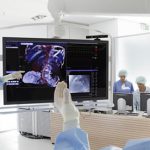
Now Siemens Healthcare has embraced this technology and developed a hands-off image manipulation system for use in the OR. The Kinect system projects an infrared grid into the room and records them using an offset camera. This allows the system to construct a 3D representation of objects in the room. The Kinect software can identify movements and objects using this data.

Siemens is using special software with the Kinect that allows it to detect and interpret fine movement of a surgeon’s hands in the operating room. The final product will allow a surgeon to browse, pan and zoom relevant patient images while they remain scrubbed and sterile, just by gesturing with their hands. This product will be tested in two hospitals in the near future.

Here’s my prediction: why will we need a big, clunky robotic system interface like DaVinci? Just have the surgeon sit in a comfortable chair, waving their hands to move the laparoscopic camera and instruments. I see especially interesting applications of this technology in military settings and in space!

Reference: Siemens Game Console Technology

New Technology: Fracture Putty

Fracture healing takes a long time, as many of our patients can attest to. Six or eight weeks, and even more may be required for full healing. Researchers at the University of Georgia and in Houston have completed an animal study on rats using a type of “fracture putty” that dramatically speeds up this process. 

The researchers used adult mesenchymal stem cells that produce a protein which is involved in bone healing and regeneration. They created a gel using these cells, and injected them into the fracture sites which were stabilized externally (imagine a rat external fixator!). The fractures healed rapidly, and within 2 weeks the rats could run and stand on their legs normally.

Bottom line: The next step is to translate this work to larger animals. Strength and durability are major concerns. The amount of stress placed on rat legs and human legs is considerably different. If this pans out, it could revolutionize fracture healing, especially in cases where there may be highly disabling segmental bone loss (read: military). It will be several years before this can move to human studies.

Reference: University of Georgia Regenerative Bioscience Center

Using A 3D Printer To Plan Orthopaedic Surgery

I’ve previously written about new printing technology applications in trauma. A recent article details a new way to use 3D printing technology for planning complex orthopaedic procedures.

An orthopedic registrar in Monklands Hospital (North Lanarkshire, Scotland) found a way to combine new printing technology and orthopaedics. CT scans are routinely taken of complex fractures. Scanners now have powerful software that enables us to create 3D reconstructions from the helical or axial images. However, these are just a series of 2D images viewed on a computer monitor.

Mr. Mark Frame found a way to convert the CT information into a format that can be used as input for a 3D printer. Using two open source (free) software packages for the Mac, OsiriX and MeshLab, he was able to create a medical quality 3D image file. The file was sent to a company that printed it using a 3D printer.

The cost? About $235 US plus a little time for a complete model of the pelvis. The advantage? The actual size 3D model can be used to select hardware and practice the repair technique. And the cost to own a 3D printer keeps coming down!

Related posts: