Tag Archives: TBI

Best Practices For TBI Patients On Oral Anticoagulants: Part 1

Over the past five years, there has been a tremendous increase in the number of patients presenting to hospitals with traumatic brain injury. The bulk of these injuries occur in the elderly, and a rapidly growing number of them are taking anticoagulants for management of their medical comorbidities. Although there is a growing body of literature addressing this issue, many practical questions remained unanswered. This is due to the lack of randomized controlled studies of the clinical problems involved. And given the ethical issues of obtaining consent for them, there likely never will be.

An interdisciplinary group of Austrian experts was convened last year to consider the most common questions asked about TBI and concomitant anticoagulant use. They reviewed the existing literature from 2007 to 2018 and combined it with their own expertise to construct some initial answers to those questions.

Over the course of my next few posts, I’ll dig into each of the questions and review their suggested answers. And remember, all these Q&A apply to patients with known/suspected TBI with known/suspected oral anticoagulant use.

Let’s start with some diagnosis questions.

Q1. Should head CT be performed in all patients with known or suspected TBI and suspected or known use of anticoagulants?

Answer: All patients with TBI and potential or known use of anticoagulants should undergo an initial screening CT scan of the head.

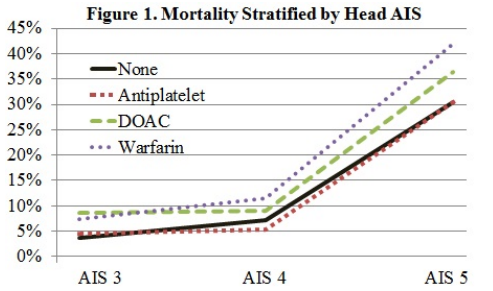
A number of systems that predict the utility of head CT already exist (e.g. Canadian head CT rules). However, they do not and cannot take into account the various permutations of drugs and other medical conditions that may influence coagulation status. Vitamin K antagonists (VKA) like warfarin have been clearly shown to increase mortality after TBI. Data involving the use of anti-platelet agents or direct oral anticoagulants (DOAC) are a bit less clear.

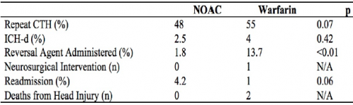
Q2. Should a repeat head CT scan be repeated in these patients, and if so, when?

Answer: Patients with intracranial hemorrhage on their initial scan should have a repeat within 6-24 hours, based on the location of the bleed.

The natural course of patients who have an identified intracranial hemorrhage is extremely unpredictable. For that reason, a repeat scan is suggested. However, there are no consistent data that would indicate when this should occur. Indications and potential for progression vary by type of bleed (subarachnoid, subdural, epidural, intraparenchymal). Thus, you must work with your neurosurgeons to arrive at a reasonable repeat interval, and it may be different for a high-risk location (epidural) vs one with low risk (subarachnoid).

Q3. Should a patient with an initial head CT that is negative be admitted for neurologic monitoring?

Answer: Patients taking only aspirin with GCS 15 and initially negative head CT may be discharged. All other patients should be admitted for at least 24 hours for neurologic monitoring as follows (q1 hr x 4 hrs, q2 hr x 8 hrs, q4 hr x 12 hrs). Repeat head CT is indicated if there is any deterioration in neurologic exam.

Multiple papers have described the occurrence of delayed intracranial hemorrhage in patients taking oral anticoagulants other than aspirin. Although some bleeds may develop days or weeks after the initial injury, the majority occur during the first 24 hours. Routine repeat head CT in this group of patients with an initially negative scan has not been found to be helpful.

Q4. What about patients with an initially negative head CT who cannot be examined neurologically (intubation, sedation, dementia)?

Answer: Unexaminable patients should undergo a repeat head CT within 6-24 hours based on the underlying risk factors for development of delayed hemorrhage.

There is no real literature on this topic, but this statement makes sense. Each center should pick a reasonable time interval and include it in their own practice guideline.

In my next post, I’ll review the panel’s recommendations on coagulation tests and target levels for reversal of the various classes of anticoagulants.

Reference: Diagnostic and therapeutic approach in adult patients with traumatic brain injury receiving oral anticoagulant therapy: an Austrian interdisciplinary consensus statement. Crit Care 23:62, 2019.

AAST 2019 #6: DOACs Part 3!

A little further down the direct oral anticoagulants (DOACs) rabbit hole please? The abstract reviewed in my last post suggested that elderly patients taking these agents actually do better than those on warfarin. So if that’s the case, do we need to be so attentive to getting followup CT scans on these patients to ensure that nothing new and unexpected is happening?

The trauma group at UCSF – East Bay performed a multi-center review of the experience at “multiple” Level I trauma centers over a three year period. They included anticoagulated patients with blunt trauma who had a negative initial head CT. Patients taking only an anti-platelet agent or a non-oral anticoagulant were excluded.  They analyzed the data for new, delayed intracranial hemorrhage, use of reversal agents, neurosurgical intervention, readmission, and death.

Here are the factoids:

  • A total of 739 records were studied: 409 on warfarin and 330 on a DOAC. Average age was 79, and half were male.
  • Repeat head CT was performed only half the time (!)
  • Delayed hemorrhage was noted in 4% of warfarin cases (9 of 224) and 2.5% of DOAC cases (4 of 159)
  • There were no interventions or deaths in the DOAC group with followup CT, or in those who did not have the repeat scan
  • There was 1 intervention in the warfarin group and two deaths attributed to TBI
  • Reversal agents were administered to 2% of DOAC patients and 14% of warfarin patients
  • The authors performed a regression analysis that showed the two strong associations with delayed hemorrhage were male sex and AIS head > 2 (!)

The authors concluded that this “largest study” suggests that DOACs “may” have a better safety profile compared to warfarin and repeat head CT is not indicated.

Now, hold on a minute!

Rule #1: No single published paper should ever change your practice. They need to be confirmed by other, hopefully better work.

Rule #2: No single abstract should make you even think about changing your practice! These are preliminary works that always need more detail, more effort, and a lot more thought. They are meant to telegraph what the authors are working on and to raise interesting questions from the audience. They should stimulate others to try to replicate and improve upon the work. In general, if something looks really good as an abstract, the next step is successful publication. This means that peers have reviewed the data and agree that it looks promising. But then it should take several years of work by the original authors and others to prove or refute the claims.

This study was small in the first place, and became smaller because half did not have repeat CT scans. The only statistically significant result was that we confirmed that the providers were not very good about getting followup scans. Just because they didn’t do it doesn’t mean it’s not indicated, especially given the nature of the data and the very small numbers.

I consider this another very small piece in the puzzle that suggests DOACs are not as evil as warfarin. There are several of these low power studies floating around right now. But we need to hunker down and really do a big study right so we can start to get a clearer picture of what we should do. For now, it’s best to treat all anticoagulants and anti-platelet agents as evil and err on the side of overtreating.

Here are my comments and questions for the presenter and authors:

  • Why was the followup head CT rate so poor? Was this a “however they like to do it” thing, was there a protocol, did the trauma centers just not believe that DOACs could be bad?
  • What were the guidelines for reversal? If the initial head CT was normal, why ever reverse? This suggests that participating centers could do whatever they wanted based on unspecified criteria.
  • Was the regression analysis helpful in any way? Being male and having a mild TBI seem rather nonspecific factors and wouldn’t help select patients for reversal or repeat scan.
  • Please provide more information on the warfarin intervention and deaths.
  • Isn’t the title of this abstract rather bold for the quality of the results presented?

I’m sure there will be some lively debate at the end of this presentation!

Reference: Repeat CT head scan is not indicated in trauma patients taking novel anticoagulation: a multi-institutional study. AAST 2019, Oral Abstract #66.

AAST 2019 #5: DOACs Part 2

In my last post, I reviewed a study that scrutinized reversal of direct oral anticoagulants (DOACs), and the outcomes of using various reversal agents. Today I’ll look at an abstract that compared in-hospital outcomes of elderly patients with severe TBI who were taking a variety of anticoagulant drugs, including DOACs.

The group at St. Joseph Mercy Hospital in Ann Arbor reviewed the dataset from the Michigan Trauma Quality Improvement Program database over a seven year period. To be included, patients needed to be at least 65 years old, suffer a fall, and have a significant head injury (AIS > 3). The final data consisted of records from 8312 patients treated at both Level I and II trauma centers across the state.

Here are the factoids:

  • 40% of patients were taking antiplatelet agents, 13% warfarin, 4% DOAC, and the remaining half or so were taking nothing.
  • The head injuries were severe, with mean AIS of 4.
  • After adjusting for “patient factors”, mortality or hospital outcomes were 1.6x more likely when warfarin was used
  • Complication risk increased 1.4x for warfarin and 1.3x for antiplatelet patients, but not for DOACs
  • Hospital length of stay was a day longer in the warfarin group (6.7 days) vs about 5.7 in the others

The authors concluded that elderly patients with severe TBI on DOACs fared better than those on warfarin. They stated that this could help alleviate concerns about DOACs in head trauma patients.

This is yet another interesting and surprising piece of the TBI on anticoagulants puzzle! It is obviously limited due to its retrospective database nature, which prevents us from asking even more interesting questions of this dataset. And it completely prevents us from looking at the specifics of each case including decision making, imaging, etc. But it’s a good start that should prompt us to find even better sources of data to tease out the details we must know in order to improve this patient group’s care.

Here are my questions for the presenter and authors:

  • I am very interested in the “patient factors” that were adjusted for to try to normalize the groups. Please describe in detail the specific ones that were used so we can understand how this influenced your results.
  • This information is intriguing, suggesting that warfarin is more evil that DOACs. What is the next step? What shall we do to further elucidate the problems, and how can we ameliorate the mortality and complication effects?

This is more good stuff about DOACs, and I can’t wait to hear the details.

Redefining Mild TBI: Who Needs To Be Transferred?

One of the more common reasons for transfer to a higher level trauma center these days is the “mild or minimal TBI.” Technically, this consists of any patient with a Glasgow Coma Scale score of 14 or 15. A transfer is typically requested for observation or neurosurgical consultation, or because the clinicians at the initial hospital are not comfortable looking after the patient.

Is this really necessary? With the number of ground level falls approaching epidemic proportions, transferring all these patients could begin to overwhelm the resources of high level trauma centers. The surgical group at Carolinas Medical Center examined their experience with a simple scoring system they designed to predict high risk minor TBI patients, and thus suitability for transfer. Here is their checklist:

Category A
  • Traumatic SAH
  • Tentorial or falcine SDH < 4mm thickn
  • Convexity SDH < 4mm thick
  • Solitary IPH < 1cm
  • Isolated intraventricular hemorrhage < 4mm
Category B
  • Any Category A lesion greater than the allowed size
  • Midline shift
  • Skull fracture
  • Compression of basal cisterns
  • Diffuse SAH or SAH involving basal cisterns
  • Subacute or chronic SDH
SAH = subarachnoid hemorrhage, SDH = subdural hemorrhage, IPH = intraparenchymal hemorrhage

Patients were considered to be low risk if they had only one or two category A lesions. If they had more than two, or any Category B lesions, they were higher risk and transfer was considered justified.

The authors retrospectively reviewed their experience with these patients over a three year period. They followed patients to see if they needed neurosurgical intervention, and evaluated the cost savings of avoiding selective transfers based on their criteria.

Here are the factoids:

  • A total of 2120 patients were studied, with 68% low risk and 32% high risk
  • Two of the low risk patients (0.14%)  ultimately required neurosurgical intervention, compared to 21% of high risk patients
  • Mean age (56), and patients taking anticoagulants or antiplatelet agents were the same in the two groups, about 2-3% for each
  • System saving by avoiding EMS transfer costs would have been $734K had the low risk patients been kept at the initial hospital

Bottom line: This study was presented as a Quick Shot paper at this year’s Eastern Association for the Surgery of Trauma meeting, so there are some key details missing. Was there an association between anticoagulation or antiplatelet agent and two failures in the low risk group? What were they, and what intervention did they require?

If this data holds up to publication, then it may provide a useful tool for deciding to keep minimal TBI patients at the local hospital. This is usually far more convenient for the patient and their family, but would require additional education of the clinicians at that hospital to help them become comfortable managing these patients. 

We use a similar tool within our Level I trauma center to decide which patients require a neurosurgical consultation. Since the low risk patients almost never require intervention, our trauma service provides comprehensive management while in hospital, and arranges for TBI clinic followup post-discharge. You can view and download a copy using the link below.

Link: Regions Hospital SAH/IPH/Skull fracture practice guideline

Reference: Redefining minimal traumatic brain injury (MTBI): a novel CT criteria to predict intervention. Quick Shot Paper #48, EAST 2019.

EAST 2019 #8: How To Keep Neurotrauma Patients At Level III Trauma Centers

Hospitals that do not have neurosurgical coverage are faced with a dilemma when they receive a head-injured patient. Do they automatically transfer to a higher level trauma center, or do they keep the patient? This is especially poignant in rural areas, where transfer times may be lengthy. If a patient doesn’t really have any significant pathology, they are likely to be evaluated at the receiving Level I or II center, then discharged all the way back home. But if they are kept at the initial hospital, there may be a nagging doubt about what happens if…

Five years ago, the group at University of Arizona – Tucson published a simple brain injury classification system that was designed to predict which injuries were likely to progress and need neurosurgical intervention. They called this system BIG for Brain Injury Guidelines. It was created and validated on a group of nearly 4,000 patients over four years, and the results have been promising. Since then, BIG has been validated using small study groups (<405) in pediatric head injury, and at Level I and III trauma centers.

Here’s the guideline:

One of the Quick Shot presentations at next week’s EAST Annual Scientific Assembly is another validation study at a Level III center in Lake Havasu City, Arizona that introduces one small modification to the guidelines. Normally, BIG is calculated after CT of the head is complete. This modification entailed BIG calculation after anticoagulation was reversed. Patients with BIG scores of 1 or two after reversal was complete were kept at the Level III, and were managed by the trauma surgeons. All BIG 3 patients were transferred to a higher level center.

Four years of trauma registry data were analyzed. During the first two years, patients with any positive BIG score were transferred. During the final two years, only patients who scored BIG 3 after reversal were moved.

Here are the factoids:

  • During the pre-BIG period, there were 72 transfers: 36 BIG-1, 23 BIG-2, and 13 BIG-3
  • Once the protocol was in place, there were 119 patients identified, 52 patients with BIG-1 or 2 who were kept and 67 BIG-3 patients who were transferred
  • 13 patients in the post-BIG time frame were excluded
  • None of the BIG-1 or 2 patients required transfer later
  • 39 of the 52 BIG-1 or 2 patients had repeat scans, and none worsened clinically, with an average hospital length of stay of 1.4 days
  • Estimated helicopter transport savings was $1.9M based on an average charge of nearly $50K per flight

The authors concluded that modified BIG could be used to triage neurotrauma patients for transfer, but cautioned that good clinical judgment should also be applied.

Here are some questions for the authors and presenter to consider in advance to help them prepare for audience questions:

  • Are you satisfied that BIG is sufficiently validated? To date, there are only a handful of validation studies and they have relatively small numbers.
  • Why did you choose to modify the score to wait until anticoagulation? Couldn’t this nullify the validation studies?
  • Do you have any practice guidelines in place to ensure consistent care of the patients you now keep at your center? Do they allow you to manage common problems like subarachnoid hemorrhage or intraparenchymal hemorrhage?
  • How did you ensure that your surgeons, hospitalists, and nurses were comfortable managing these neurotrauma patients? Did you have any educational sessions or other training for things like GCS monitoring and neuro exam?
  • How do you reverse anticoagulation, and how long does that usually take? Plasma and prothrombin complex concentrate are commonly used, but with vastly different reversal times. And what do you do about aspirin, clopidogrel, and the novel oral anticoagulants?
  • Why did you exclude 13 patients once you started using BIG?
  • Has your hospital administration provided any numbers regarding increased revenue from this practice? Your hospital is larger (171 beds), but this type of information will be vital for small, critical access hospitals.

This is very interesting work, and highly applicable to rural trauma centers!

References:

  • Successful management of select radiographic intracranial injuries in a rural trauma center without neurosurgeon coverage using a modified brain injury guideline. EAST 2019, Quick Shot Paper #6.
  • The BIG (brain injury guidelines) project: Defining the management of traumatic brain injury by acute care surgeons. J Trauma 76(4):965-969, 2014.