Tag Archives: spleen

Does Trauma Center Level Make A Difference In Treating Solid Organ Injury?

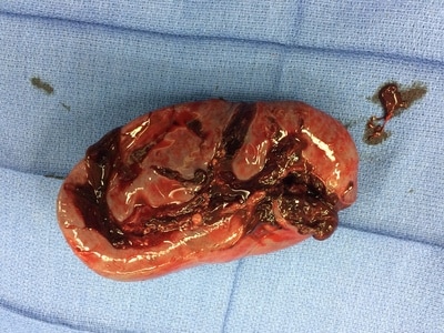
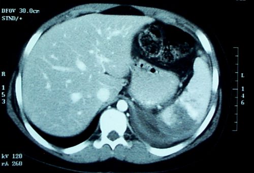
In the last two posts, I reviewed contrast anomalies in solid organs, specifically the spleen. Today, I’ll be more general and examine a recent paper that compared management and outcomes after the other major solid organ injury, liver, at Level I vs Level II trauma centers.

There are several papers that have detailed overall differences in outcomes, and specifically mortality, at Level I and II centers. Some of these show outcomes that are not quite as good at Level II centers when compared to Level I. On paper, it looks like these two levels should be very similar. Take away research and residents, and maybe a few of the more esoteric capabilities like reimplantation, and aren’t they about the same?

Well, not really. They can be, though. Level I criteria are fairly strict, and the variability between difference Level I centers is not very great. Level II criteria are a bit looser, and this allows more variability. Many Level II centers function very much like a Level I, but a few are only a bit higher functioning than a Level III with a few extra surgical specialists added in.

A paper currently in press used the Michigan Trauma Quality Improvement Program (MTQIP) data from all 29 ACS verified Level I and II centers in the state (wow!). Six years of information was collected, including the usual demographics, outcome data, and management. A total of 538 patients met inclusion criteria, and this was narrowed down to 454 so statistical comparisons of similar patients could be made for Level I vs Level II centers.

Here are the factoids:

  • Mortality was significantly higher in Level II centers compared to Level I (15% vs 9%) and patients were more likely to die in the first two days, suggesting hemorrhage as the cause
  • Patients were more likely to die in the ED at Level II centers, despite a significantly lower Injury Severity Score (ISS)
  • Pneumonia and ARDS were significantly more likely to develop in Level II center patients
  • Level II centers used angiography less often and took patients to the OR more frequently
  • Level II centers admitted fewer patients to the ICU, but ICU admission was associated with significantly decreased mortality
  • Complications were fewer at Level II centers, but they were less likely to rescue patients when they occurred

Bottom line: Level I and II centers are supposed to be roughly the same, at least on paper. But a number of studies have suggested that there are more disparities than we think. Although this paper is a retrospective review, the sheer number of significant differences and its focus on one particular injury makes it more compelling.

So what to do? Tighten up the ACS Orange Book criteria? That’s a slow and deliberate process that won’t help our patients now. The quickest and most effective solution is for all centers to adopt uniform practice guidelines so they all perform like the highly successful Level I programs in the study. There are plenty of them around. If you are not yet using one, I urge you to have a look at the example below. Tweak it to fit your center. And use your PI program to trend the outcomes!

Related post:

Reference: Variability in Management of Blunt Liver Trauma and Contribution of Level of ACS-COT Verification Status on Mortality. J Trauma, in press, Dec 1, 2017.

Natural History of the Splenic Blush

In my last post, I described the two types of solid organ “blushes.” I also described my thoughts on the natural history of these findings. Now, a multicenter study on the natural progression of the splenic “blush” has just been published. I found this paper very interesting, because it challenged some of my own existing beliefs. But once I read it, my enthusiasm faded.

The Western Trauma Association sponsored a multicenter (17 Level I and II centers) review of data collected prospectively over an unspecified period of time. Patients were excluded if their injury was older than 24 hours, if they had a previous splenic injury, and if they had any number of diseases or hereditary conditions that might affect the spleen. Strict definitions of nonbleeding and actively bleeding injuries were applied, and detailed information on intervention and outcomes was collected.

Here are the factoids:

  • 200 patients were enrolled from 17 centers, but the paper does not state how long that took
  • 20% were low grade (1 or 2) and 80 % high grade (3-5)
  • 29% had a pseudoaneurysm, and 83% showed extravasation, which means that several patients had both
  • 15% underwent early splenectomy, 59% underwent angiography, and 26% were observed
  • For those with initial angiography, 6% had repeat angio and 7% eventually underwent splenectomy
  • Of those were were initially observed, 9% had delayed angio and 8% underwent splenectomy
  • Based on a read by an expert radiologist, an actively bleeding injury was associated with a 41% splenectomy rate
  • The authors conclude that the majority of patients with spleen injury with pseudoaneurysm or extravasation are managed with angio and embolization and that splenectomy remains a rare event (??)

Bottom line: This paper just doesn’t do it for me. The biggest problem is that it is what I call a “we do it the way we do it” study. It examines how 17 different centers evaluate and treat patients with significant splenic injury. There was no guidance or guideline on how to treat, so they each did it their way. And the number of patients was small.

They don’t tell us anything about the use or effectiveness of angio by grade. Or whether the specific hospitals routinely rely on angio rather than just going to the OR for high grade injuries (typically if angio response times are long).

Unfortunately, this paper gives the appearance of containing a lot of interesting stuff. But a 15% initial splenectomy rate is not a “rare event” in my book. Everything published here is at odds with what I’ve observed over the years for centers with well developed management guidelines and easy access to angio (< 5% splenectomy rate in hemodynamically stable patients with nonoperative management).

My recommendation is to send all stable patients with pseudoaneursym and/or extravasation to angio immediately! Yes, some will have nothing found by the time they get to angio, and you’ll have to come up with a plan at that point. But most have something wrong, and it won’t stop until it’s been plugged up (or your patient bleeds to death, whichever comes first)!

This article has all the right buzzwords: multicenter, prospective data, etc. But it’s already been moved to my recycle bin. 

Related post:

Reference: Natural history of splenic vascular abnormalities after blunt injury: A Western Trauma Association multicenter trial. J Trauma 83(6):999-1005, 2017.

Splenic Vascular Blush

Contrast blush is always a concern when seen on CT of the abdomen for trauma. It can represent one of two things, and both are bad:

  • Active extravasation of contrast
  • Splenic pseudoaneurysm

These two clinical issues can be distinguished by looking at the location of the contrast and its persistence. A pseudoaneurysm is located within the parenchyma, and the contrast will wash away, so it will not be visible on delayed images. Contrast that extends beyond the parenchyma or persists in delayed views represents active bleeding. In either case, the failure rate of nonoperative management exceeds 80% in adults without additional measures being taken.

Clinically, these patients usually act as if they are losing volume and require additional crystalloid and/or blood transfusion. The natural history in adults is for bleeding to continue or for the pseudoaneurysm to rupture, resulting in a quick trip to the operating room.

If vital signs can be maintained with fluids and blood, a trip to interventional radiology may solve the problem. Selective or nonselective embolization can be carried out and patients with only a few bleeding points can be spared operation. However, if multiple bleeding areas are seen, it is probably better to head to the OR for splenorrhaphy or splenectomy.

The image below shows likely areas of extravasation. They are a bit large to be pseudoaneurysms.

Spleen Blush-CT

Children are different than adults. Extravasation from spleen injuries in prepubescent children frequently stops on its own. Angiography should only be used if the child is failing nonoperative management.

Next post: A new paper looks at the natural history of these lesions.

Spleen Embolization In Adolescents?

Modern day nonoperative management of solid organ injury in adults came to be due to its success rate in children. But if you look at the practice guidelines for adults, they frequently include a path for angioembolization in certain patients. In children, embolization is almost never recommended.

But what about that gray zone where children transition to adults? How young is too young to embolize? Or how old is too old not to consider it?

The adult and pediatric trauma groups at Wake Forest looked at this question by reviewing their respective trauma registry data. They looked specifically at patients age 13-18 who presented with a blunt splenic injury over a 8.5 year period. About halfway through this period, adult patients (> 16 years) were sent for embolization not only for pseudoaneurysm or extravasation, but also for high grade injury (> grade 3).  Patients under age 16 were managed by the pediatric trauma team, and those 16 and older by the adult team.

Here are the factoids:

  • Of the 133 patients studied, 59 were “adolescents” (age 13-15) and 74 were “adults” (16 or older)
  • Patients managed by the adult team sent 27 of their 74 patients for angiography
  • Those managed by the pediatric team were never sent to angiography
  • The failure rate for nonoperative management was statistically identical, about 4% in adults and 0% in adolescents
  • For high grade injuries, the adult team sent 27 of 34 patients to IR, whereas the pediatric team sent none of 36. Once again, failure rate was identical.

Bottom line: We already know that too many adult trauma centers send too many younger patients to angiography for solid organ injury. This study tries to tease out when a child becomes an adult, and therefore when angiography should begin to be considered. And basically, it showed that through age 15, they can still be considered as and treated like children, without angiography.

But remember, these numbers are relatively small, so take this work with a grain of salt. If you are managing a younger patient nonoperatively, and they continue to show evidence of blood loss (ongoing fluid/blood requirements, increasing heart rate), angiography may be helpful in avoiding laparotomy as long as your patient remains hemodynamically stable. But consult with your friendly neighborhood pediatric surgeon first.

Related posts:

Reference: The Spleen Not Taken: Differences in management and outcomes of blunt splenic injuries in teenagers cared for by adult and pediatric trauma teams in a single institution. J Trauma, in press, May 2017.