Tag Archives: spine

What Are: These Spondylo… Words

Spondylosis. Spondylolisthesis. Spondylitis. These words are tossed about blithely by our orthopedic and neurosurgical spine colleagues. But many trauma professionals are confused by the terms. What do they mean? What do they look like?

Let’s start with the root of the word, spondylo… This part is derived from the Greek word spondylos, meaning spine. Now let’s combine it with some of the usual suffixes.

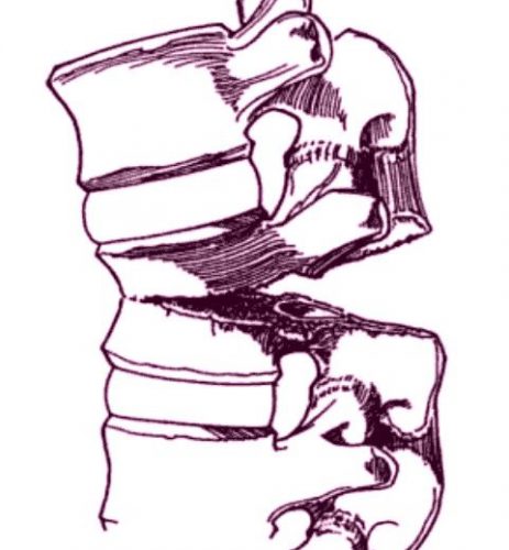
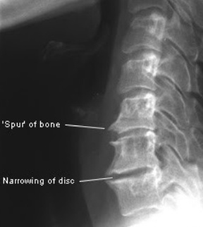
The first one is -osis, so this creates the word spondylosis. Although -osis can denote the “condition of being a …”, in medicine it frequently means a disease or pathological process. Think diverticulosis of the colon. Spondylosis usually denotes a degenerative process of the spine. This is typically due to arthritis and results in bone spurs and disc narrowing. Here is an image of a spine with significant spondylosis:

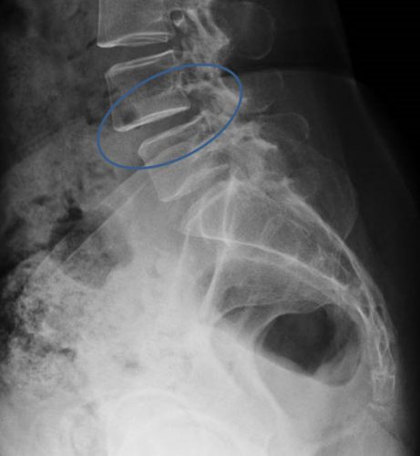
Now let’s add -listhesis. This is another Greek word that means “slipping or falling.” So in this case, the full word means one vertebra slipping over another. Here’s an image of an anterior spondylolisthesis:

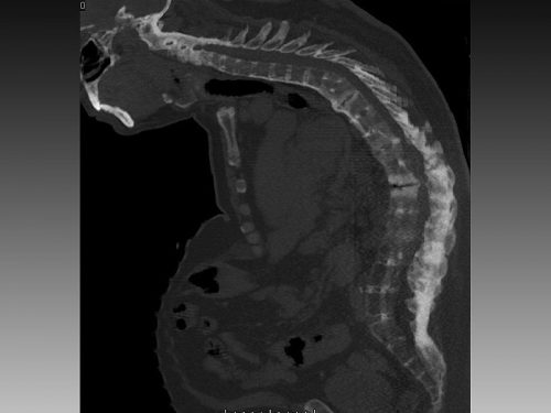
Finally, let’s add -itis. This is the Greek suffix for inflammation. So spondylitis is an inflammatory process of the spine. This can be due to infectious or autoimmune causes. One of the more common types is ankylosis spondylitis, which is an autoimmune variant of rheumatoid arthritis. This causes inflammation of the facet joints and the stabilizing ligaments, leading to fused vertebra and a characteristic patient posture. Here’s a rather extreme case:

I hope this little vocabulary lesson has been helpful. Now go impress your spine specialty colleagues!

Bullet In The Disk Space: Big Deal Or Not?

In an earlier post, I reviewed the problems with lead poisoning that can occur if a bullet remains in contact with a joint space / synovial fluid, or ends up in the GI tract. But what about if it comes to rest in an intervertebral joint space? They’re dry, right?

The first case report I could find dates back to 1981. A male presented to Parkland Memorial Hospital 12 years after a gunshot to the abdomen in which the bullet lodged in a disk space. He was treated for a GI bleed, but was also noted to have many signs and symptoms of high lead levels. These included irritability, anemia, headache, lethargy, muscle weakness and confusion. A blue line was noted on the gums. X-ray of the lumbar spine showed the bullet fragment in the center of the disk space, and a cystic mass in the prevertebral area that appeared radiodense as well. Blood lead levels were elevated. The patient underwent diskectomy, resection of the mass, chelation therapy, and recovered.

Another case report from 2010 was similar in many ways. The patient was young, had a gunshot 5 years previously, and presented with symptoms of lead poisoning. The appearance of the bullet in the disk space was similar to the last case, in that the bullet could be seen within it, and there appeared to be additional radiopaque material surrounding it. It almost looked like lead was flowing out of the bullet into the disk. This case was also treated with surgical removal and chelation with a successful result.

A literature review was conducted 15 years ago that examined other case reports of bullets in the spine. Over a 25-year period 238 patients were identified with this injury. Only 12 had bullets or fragments in the disk space. All were tested for plumbism, and only one was positive. He underwent diskectomy and resection with resolution of the high lead levels.

Bottom line: We know that a bullet in contact with synovial fluid is bad, with rapid leaching of lead into the circulation. There are also suggestions that lead in contact with CSF can cause a similar problem. However, the intervertebral disk space is usually considered to be “dry” and doesn’t usually cause a problem.

However, patients with a bullet in this location should be cautioned that they do have a small risk of developing lead poisoning. They should be tested about six months post-injury to see if lead levels are on the rise. They should also be cautioned to report the development of new back pain. Structural disruption by the bullet may slowly lead to anatomic changes that result in chronic pain. And be very suspicious if there is radiopaque material in the disk space in addition to the bullet itself!

References:

  1. Acute lead intoxication from a bullet in the intervertebral disk space. JBJS 63A(7):1180-1182, 1981.
  2. Lead Poisoning by Intradiscal Firearm Bullet. Spine 35(4):E140-E143, 2010.
  3. Long-Term Clinical Manifestations of Retained Bullet
    Fragments Within the Intervertebral Disk Space. J Spinal Disord Tech 17(2):108-111, 2004.


ACS Trauma Abstracts #4: Timing Of DVT Prophylaxis In Spine Trauma

Spine trauma is one of the high-risk indicators for deep venous thrombosis (DVT). Unfortunately, there is a great deal of variability in the start time for chemical prophylaxis for this injury, especially after the patient has undergone surgery. In part, this is due to a lack of good literature and guidelines, and in part due to the preferences of the spine surgeons who operate  on these patients.

A group at the University of Arizona in Tucson performed a large database review (looks like National Trauma Databank, although they don’t say in the abstract) looking at “early” vs “late” administration of prophylaxis after surgery in these patients. The spine injury was the predominant one, with all other systems having an abbreviated injury score (AIS) < 3. They matched two years worth of patients for demographics, initial vitals, type of operative intervention, and type of heparin to assess the impact of prophylaxis timing.

Here are the factoids:

  • Nearly 40,000 patient records were reviewed, and over 9,500 met the spine injury criteria with operation and prophylaxis. A total of 3,556 could be matched for analysis.
  • These patients were split in half for matching, late (>48 hrs) versus early (<48 hrs)
  • DVT rate was significantly lowe in the early prophylaxis group (2% vs 11%)
  • PE rate and mortality were the same between groups
  • Return to OR and blood transfusion rates were identical (1% and 1-2 units)

Bottom line: Once again, we see that “early” prophylaxis for DVT is probably desirable and mostly harmless, even after a spine operation. Many surgeons still have an irrational fear of giving heparin products in patients who have some risk of bleeding. The body of literature that supports early use just keeps growing. One observation, though: as in most other studies, pretty much whatever we do for DVT has a negligible impact on PE and mortality. We can only treat the clots, but not their major aftermath.

Reference:  Optimal timing of initiation of thromboprophylaxis in spinal trauma after operative intervention: – propensity-matched analysis. JACS 225(4S1):S59-S69, 2017.

Physical Exam And Thoracolumbar Spine Fractures

The physical exam is an important part of the initial evaluation of trauma patients. Sometimes it actually makes the diagnosis, but much of the time it focuses further studies like x-rays or lab tests. But we can also use it as a tool to avoid further imaging. For example, consider clinical clearance of the cervical spine. A negative exam in a reliable patient allows us to remove the cervical collar.

Can we apply the same thinking to the thoracic and lumbar spines? Many of us do. No pain or tenderness equates to no imaging or log-roll precautions.

The trauma group at LAC+USC looked at this one a few years ago. They studied every blunt trauma patient over a 6-month period, and first determined if they were “evaluable.” This meant not intoxicated, head injured (GCS<15), and no distracting injury (determined very subjectively). All underwent a standard exam of the TL spine by a resident or attending surgeon.

Here are the factoids:

  • 886 patients were enrolled, and 218 (25%) were not evaluable using the criteria above
  • 11% of the non-evaluable patients were found to have a TL spine fracture by CT, whereas only 8% of the evaluable group did
  • Of the evaluable patients, half (29) had no signs or symptoms of fracture
  • Of those 29 without signs or symptoms, two had a “clinically significant” fracture. Both were younger (20 and 59). One had a T7 compression and a transverse process fracture, the other a T9 compression fracture. Both were treated with a TLSO brace.
  • Of the 27 who could not be examined, 11 had “clinically significant” fractures; 8 were treated with TLSO and 6 with surgery (obviously some overlap there)

Bottom line: So physical exam of the thoracic and lumbar spine sucks, right? Not quite so fast! First, this is a small-ish study, but with enough patients to be intriguing. The biggest issue is that we don’t really know what is “clinically significant.” Treatment of stable fractures of the spine is controversial, and our friendly neighborhood neurosurgeons vary tremendously in how they do it. Ask five neurosurgeons and you’ll get six different answers.

Braces are expensive, and the optimal choice is not clear yet. At my hospital, we are treating select ones with a binder for comfort or a simple backpack brace. The fancier ones like the TLSO easily cost over $1000!

At this point, I recommend that you use a good blunt imaging practice guideline like the one below, coupled with a good physical exam. If the patient has sufficient mechanism to break something (which decreases with patient age), then image them. If they don’t, but have an abnormal exam, image them anyway. And we’ll wait for the next bigger/better study!

Related posts:

Reference: Clinical examination is insufficient to rule out thoracolumbar spine injuries. J Trauma 70(1):174-179, 2011.

The Chance Fracture

Centers that take care of blunt trauma are familiar with the spectrum of injury that is directly attributable to seat belt use. Although proper restraint significantly decreases mortality and serious head injury, seat belts can cause visceral injury, especially to small bowel.

Lap belt use has been associated with Chance fracture (flexion distraction injury to the lumbar spine) since 1982. The association between seat belts and intra-abdominal injury, especially with an obvious “seat belt sign” was first described in 1987.

chance-fracture-21

Chance fracture. The vertebra appears to split in half from posterior to anterior.

Twenty years ago, orthopedic surgeons in Manitoba finally put two and two together and reported a series of 7 cases of Chance fractures. They noted that 6 of the fractures were associated with restraint use. Seat belt sign was also present in 5 of the 6 patients with fractures and three of the six had bowel injuries.

The authors noted that many provinces were mandating seatbelt use at the time, and they predicted that the number of Chance fractures, seat belt signs and hollow viscus injuries would increase. On the positive side, the number of deaths and serious head injuries would be expected to decline.

Although this was a small series, it finally cemented the unusual Chance fracture, seat belt sign, and bowel injury after motor vehicle trauma.

Thankfully, three point restraints (lap belt + shoulder harness) has been required in the seats next to doors for a long time. And since 2007, they have been mandated in the middle seat as well. Thus, these injuries seldom occur in any but the oldest (beater) cars on the road. They are seen more frequently now with sports and extreme sports injuries.

Chance fractures are frequently unstable, involving all three columns of the spine. The anterior column fails under compression, and the middle and posterior columns fail from the distraction mechanism. Usually, this fracture pattern requires operative fixation. However, if the posterior column is intact, a TLSO brace can be tried. This fracture is at risk for non-union and development of kyphosis or a flat back, which can lead to chronic pain and an abnormal posture.

Reference: Pediatric Chance Fractures: Association with Intra-abdominal Injuries and Seatbelt Use. Reid et al. J Trauma 30(4) 384-91, 1990.