Tag Archives: spinal cord injury

Best of EAST 2024 #1: MAP And Spinal Cord Injury

The use of elevated mean arterial pressure (MAP) to help manage spinal cord injury has been a mainstay of treatment for years. The concept is similar to that used for management of severe traumatic brain injury. The theory is that there may be areas of the brain that are damaged, but not irretrievably so. Increasing MAP should improve perfusion and may protect areas in jeopardy from secondary injury.

As with so much in neurotrauma, few large and/or prospective studies exist. Although most centers have specific algorithms and MAP goals, optimal treatment still needs to be determined.

EAST sponsored a prospective, multicenter study to identify factors influencing neurologic outcomes after spinal cord injury. MAP augmentation was monitored, specifically its impact on the American Spinal Injury Association (ASIA) score between admission and discharge.

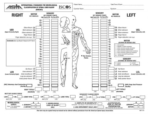
The ASIA Score is calculated by performing a very detailed exam consisting of myotomal motor function, a dermatomal sensory exam, and an anorectal exam. The exam takes quite some time to complete. The copy of the worksheet below should give you an idea of the level of detail:
The study was performed over 20 months, and 19 centers participated. They entered 222 patients, but only 164 had pre- and post-ASIA scores for comparison.

Here are the factoids:

  • Of the 164 patients studied, only 36 improved vs. 128 that showed no improvement by ASIA score
  • Demographics, hospital and ICU length of stay, and mortality were not significantly different between the groups
  • ISS was nearly identical (23 vs 25)
  • Three-quarters of injuries were to the cervical spine, about 10% to the lumbar spine, and the remainder to the thoracic spine. There was no correlation between injury location and recovery.
  • Presentation in the trauma bay (blood pressure, pulse, MAP, lactate, and Hgb) were the same in both groups
  • The MAP goal of >85 mm Hg was met about 75% of the time in both groups
  • Duration of MAP therapy was the same for the two groups, from 99-113 hours
  • There was a trend toward increased cardiac issues (atrial fibrillation, v-tach, elevated troponin) in the group with improved spinal cord recovery. This may be due to the medications used to increase MAP.

Bottom line: This is very interesting work and will make us question the utility of MAP therapy for spinal cord injury. However, this is not a cut-and-dried conclusion. Here are several things that come to mind:

  • What was the definition of “improvement?” ASIA is a complicated scoring system with many steps in the evaluation. Usually, the results are condensed into an overall “ASIA Impairment Scale,” or AIS.
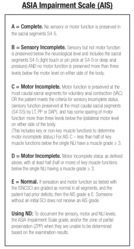
    The AIS is not very granular, meaning that each step in the scale represents a large difference in function. Could patients have had improvements that did not change the AIS score but were functionally significant for the patient? For example, an improvement from a C5 to a C6 level makes a big difference in daily activities.
  • Was the study large enough? It is difficult to accumulate a large series of spinal cord injury patients. Combining this point with the previous one, was the statistical power present even to detect a meaningful difference in the AIS?
  • Was MAP>85 torr maintained reliably and for long enough? Patients had MAP therapy for just over four days, and it was only maintained above the threshold about 75% of the time. We have good evidence in the brain injury literature that a single bout of hypotension in patients with severe TBI significantly increases mortality. Could it be that maintaining increased spinal cord perfusion is equally important? Could a single low MAP cause damage? It would be interesting to see if patients who had very consistent MAP therapy, say greater than 90% or 95% of the time, had any difference in outcomes. Unfortunately, I suspect that the numbers would be far too low to prove anything.

This abstract brings up some interesting questions. However, I would not consider throwing out the use of MAP goals based on it. We need more patients to study and be better at applying this treatment if we hope to uncover whether it really works.

Reference: Does mean arterial pressure augmentation improve neurological recovery of blunt spinal cord injuries: an EAST multicenter trial. EAST 2024 Podium paper #1.

Spinal Cord Concussion In Student Athletes

Spinal cord injuries are typically devastating injuries with profound consequences for function and life expectancy. However, a small percentage result in rapidly reversible symptoms. Because these temporary injuries are rare, they tend to cause confusion among clinicians.

Technically, a spinal cord concussion (a “zinger” or “stinger” is an example) is a mild cord injury that results in transient neurologic disturbances. The deficits can be sensory, motor or both, and typically resolve in less than 48 hours. The injuries tend to involve the mid-portion of the cervical cord or the cervico-thoracic junction, since these are the areas of maximum mobility. In a few cases, the athlete has congenital narrowing of the spinal canal which predisposes them to injury. In most cases, the injury probably occurs due to the flexibility of the young spine.

The usual management consists of an MRI of the spine followed by admission and frequent neurologic checks to ensure ongoing resolution. MRI is typically negative in a true concussion. If a signal change is seen, then technically a cord contusion is present. Management is the same for both. There is no indication to give steroids. Evaluation of the ligaments is critical to determine if a collar will be necessary.

Recovery is rapid and complete. But what is the answer to the inevitable question, “when can he/she return to play?” In adult players, the literature suggests that it may be safe to return once they have fully recovered. There is little guidance for kids.

Here’s what I tell the parents: This event has shown that, given the right force applied to your child’s neck, the bones can move enough to injure their spinal cord. This time, the cord was just tickled a little bit. But if the bones had moved just another millimeter or two, this injury could have been permanent and they would never have walked again. I recommend that they do not play this sport again.

Some of you may disagree. I’d be very interested in hearing your comments. 

Reference:

  • First mention: About concussion of the spinal cord. Wein Med Jahrb 34:531, 1879.

Trauma Mythbusters: Spinal Cord Injury From Airway Management

Airway management is one of the most anxiety provoking procedures performed by trauma professionals. The main fear is that the airway will be lost during attempts to secure it and patient demise will soon follow. Add some facial fractures, bleeding, and an “unfavorable body habitus”, and the average prehospital or ED provider is really on edge.

The next most common fear is that providing a definitive airway in a patient with a known (or even suspected) cervical spine injury could cause a catastrophic neurologic injury. This was first addressed back in 1989 (before the time of video laryngoscopy, and when flexible scopes were rarely available in the ED). The authors found no verifiable reports of such an injury in the entire English literature

Over the years, a few case reports have cropped up. As in so much of the medical literature, causality is hard to prove. The patient was normal before anesthesia, and afterwards they were not. Had to be the intubation, right?

Not so fast! Let’s break it down and look at what we do know. Biomechanical studies have shown that the manipulation that occurs in direct laryngoscopy isn’t as bad as it looks. Studies in uninjured models are enlightening (minimal movement with blade insertion, slight rotational movements with blade elevation, and a little more rotation during the intubation). Most of this (slight) movement occurs from occiput to C2, with little motion at all at C3 and below.

But that was on an uninjured model. What about ones that simulate an injured spine? Specifically an injury in the upper spine area that we know moves?

  • Cricoid pressure caused no appreciable changes in the spine
  • Chin lift and jaw thrust reduced space available for the cord (SAC) by 1 and 2.5mm, respectively, and caused an angulation of about 4-5 degrees
  • SAC narrowed by only 1.5mm, even with maximum flexion and extension
  • Oral and nasal intubation narrowed SAC by 1.6mm, and resulted in a maximum of 2.5 degrees of rotation
  • Video laryngoscopy results in about half of the rotational movement of direct laryngoscopy
But what about these sporadic reports of neurologic deficits after intubation? What is often neglected is that spinal blood flow and long-term neck positioning have a major impact on cord function. Even relatively mild malpositioning of the cervical spine for extended periods during an OR case have been documented. 

Bottom line: From a mechanical standpoint, even in unstable spine models, the maneuvers we use in preparation for intubation cause more movement of the spine than does the intubation procedure itself. The true number of spinal cord injuries actually (and provably) caused by intubation approaches zero. The literature suggests that video laryngoscopy results in less overall movement during intubation, but it doesn’t seem to have an impact on cord injury (you can’t get less than zero). 

References:

  • Spinal cord injury and direct laryngoscopy – the legend lives on. Br J Anesth 84(6):705-709.
  • Airway management in adults after cervical spine trauma. Anesthesiology 104(6):1293-1318, 2006.

Spinal Cord Concussion In Student Athletes

Spinal cord injuries are typically devastating injuries with profound consequences for function and life expectancy. However, a small percentage result in rapidly reversible symptoms. Because these temporary injuries are rare, they tend to cause confusion among clinicians.

Technically, a spinal cord concussion (a “zinger” or “stinger” is an example) is a mild cord injury that results in transient neurologic disturbances. The deficits can be sensory, motor or both, and typically resolve in less than 48 hours. The injuries tend to involve the mid-portion of the cervical cord or the cervico-thoracic junction, since these are the areas of maximum mobility. In a few cases, the athlete has congenital narrowing of the spinal canal which predisposes them to injury. In most cases, the injury probably occurs due to the flexibility of the young spine.

The usual management consists of an MRI of the spine followed by admission and frequent neurologic checks to ensure ongoing resolution. MRI is typically negative in a true concussion. If a signal change is seen, then technically a cord contusion is present. Management is the same for both. There is no indication to give steroids. Evaluation of the ligaments is critical to determine if a collar will be necessary.

Recovery is rapid and complete. But what is the answer to the inevitable question, “when can he/she return to play?” In adult players, the literature suggests that it may be safe to return once they have fully recovered. There is little guidance for kids.

Here’s what I tell the parents: This event has shown that, given the right force applied to your child’s neck, the bones can move enough to injure their spinal cord. This time, the cord was just tickled a little bit. But if the bones had moved just another millimeter or two, this injury could have been permanent and they would never have walked again. I recommend that they do not play this sport again.

Some of you may disagree. I’d be very interested in hearing your comments. 

Reference:

  • First mention: About concussion of the spinal cord. Wein Med Jahrb 34:531, 1879.

DVT: Does spinal cord level make a difference?

Deep venous thrombosis (DVT) is always a concern in trauma patients. Patients with spine and spinal cord injury have been shown to be at higher risk for DVT than many other trauma patients, with a reported incidence ranging from 5% to 70%. However, a few studies have suggested that paraplegics are actually at higher risk than quadriplegics. This just doesn’t seem to make sense.

A NTDB study was done to look at this issue. A total of 18,000+ patients were reviewed, and correlations with spinal cord injury level, demographics, comorbidities and associated injuries were determined.

High cervical (C1-4) and lumbar cord injuries had the lowest DVT rates at about 3%. Lower cervical (C5-7) and high thoracic (T1-6) had the highest rates at 5% and 6.3%, respectively. The lower thoracic spine was about 4.5%. These differences were statistically significant, and the authors also confirmed the usual DVT suspects as being significant (increasing age, increasing injury severity, TBI, chest trauma, and male gender).

Bottom line: Yes, this study does confirm the suspicion that paraplegics are at higher risk for DVT than quadriplegics. Why? We don’t know. And although it is statistically significant, is it clinically significant? I’m not so sure. We’re talking another 1-2 spinal cord injured patients with DVT for every 100 quadriplegics treated. How many do you admit per year? At my institution, this means that there will be 1 additional DVT in this patient group every three to four years. It’s hard to justify making any changes to existing protocols based on these new facts. Always look at the practical side of what you read!

Related posts:

Reference: Risk of venous thromboembolism after spinal cord injury: not all levels are the same. J Trauma 71(5):1241-1245, 2011.