Tag Archives: repeat

Best Of EAST #9: Routine Repeat Head CT For TBI Patients On Antithrombotic Agents

The data we use as guidance for repeat head CT in elderly patients who sustain mild TBI while taking antithrombotic therapy remains limited. There is a slowly growing consensus that the need is limited, but there is still a very wide variation in practice patterns.

The group at HCA Healthcare Nashville collected data from 24 system hospitals on this very specific cohort of patients: elderly (age > 55), head trauma with GCS 14-15, an initial head CT, and no other injuries with AIS > 2. They divided these patients into two groups based on whether they were currently taking antithrombotic (AT) therapy. Rate of delayed intracranial hemorrhage (ICH), need for neurosurgical intervention, and mortality were compared.

Here are the factoids:

  • About 3,000 patients were enrolled and only 10% had a repeat head CT
  • Of those who were rescanned, 10% of patients on meds had a new ICH vs 6% in those not taking meds (not statistically significant)
  • Extrapolating those numbers to all patients, the rate of delayed ICH would be 0.7% in patients not taking AT vs 1.0% for those who were (also not significant)
  • Mortality attributable to a head bleed occurred in only one patient who was made comfort care
  • There were no neurosurgical procedures performed in either group

The authors concluded that this specific subset of patients has a very low rate of delayed ICH, and that there are minimal clinical consequences in those that do. They do not support repeat head CT.

Bottom line: This abstract adds to the growing body of literature that shows little benefit to repeat head CT scan after a negative initial study, even if the patient is on blood thinners. Many previous studies involve only a single center and/or have smaller numbers. This one is larger because of the size of the HCA trauma system, and answers a simple set of questions on a limited subgroup of patients: elderly, mild TBI, with limited other injuries.

My back of the envelope power calculations show the authors may be a little short of the number of subjects to be able to show that the difference in the number of delayed ICH (0.7% vs 1.0%) is statistically significant. But the numbers are close enough and the p value so large (0.3) that they are probably right. This is completely offset by the absence of necessary neurosurgical interventions and the single attributable death.

Many trauma centers, including my own, have adopted a “no repeat scan” policy after a negative initial scan, even on thinners. In fact, unless the patient has some other injury that requires admission, they are discharged home with a responsible adult.

Here are my questions for the authors and presenter:

  • Did you do any type of power analysis to determine if the large number of patients included was actually large enough?
  • The term “antithrombotic therapy” is used broadly; which agents were considered in this category? Traditional warfarin therapy? Aspirin and other antiplatelet agents? DOACS?
  • Have you changed your system guidelines to reflect your work?

This is important and practical work! I’m looking forward to hearing all the details.

Reference: ROUTINE REPEAT BRAIN CT SCANNING IS UNNECESSARY IN OLDER PATIENTS WITH GCS 14-15 AND A NORMAL INITIAL BRAIN CT SCAN REGARDLESS OF PREINJURY ANTITHROMBOTIC USE: A MULTICENTER STUDY OF 3033 PATIENTS. EAST 35th ASA, oral abstract #31.

Don’t Repeat Trauma Activation X-rays!!

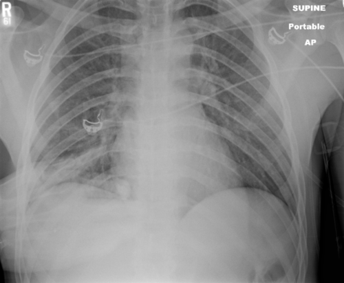
You are in the middle of a fast-paced trauma activation. The patient is awake, and mostly cooperative. The x-ray plate is under the patient and everyone stands back as the tech gets ready to fire the x-ray machine. At that very moment, your patient reaches up and places his hand on his chest. Or one of the nurses reaches over to check an IV site.

The x-ray tech swears, and offers to re-shoot the image. What do you do? Is it really ruined? They have an extra plate in hand and are ready to slide it under the patient bed.

The decision tree on this one is very simple. There are two factors in play: what do you need to see, and how hard is it to see? The natural reaction is to discard the original image and immediately get a new one. It’s so easy! Plus, the techs will take heat from the radiologist because of the suboptimal image. But take a look at this example of a “ruined” chest xray.

It’s just the patient’s hand! You can still see everything that you really need to.

Bottom line: You are looking for 2 main things on the chest x-ray: big air and big blood. Only those will change your management in the trauma bay. And they are very easy to see. Couple that with the fact that an arm overlying the image does not add a lot of “noise” to the image. So look at the processed image first. 99% of the time, you can see what you need, and will almost never have to repeat. [Hint: the same holds true for the pelvic x-ray, too. You are mainly looking for significant bony displacements, which are also easy to see.]

Related posts:

Do You Really Need To Repeat That Xray?

It happens all the time. You get that initial chest and/or pelvic xray in the resuscitation room while evaluating a blunt trauma patient. A few minutes later the tech returns with another armful of xray plates to repeat them. Why? The patient was not centered properly and part of the image is clipped.

Do you really need to go through the process of setting up again, moving the xray unit in, watching people run out of the room (if they are not wearing lead, and see my post below about how much radiation they are really exposed to), and shooting another image? The answer to the question lies in what you are looking for. Let’s address the two most common (and really the only necessary) images needed during early resuscitation of blunt trauma.

First, the chest xray. You are really looking for 3 things:

  • Big air (pneumothorax)
  • Big blood (hemothorax)
  • Big mediastinum (hinting at aortic injury)

Look at the clipped xray above. A portion of the left chest wall is off the image. If there were a large pneumothorax on the left, would you be able to see it? What about a large hemothorax? And the mediastinum is fully included, so no problem there. So in this case, no need to repeat immediately.

The same thing goes for the pelvis. You are looking for gross disruption of the pelvic ring, especially posteriorly because this will cause you to intervene in the ED (order blood, consider wrapping the pelvis). So if parts of the edges or top and bottom are clipped, no big deal.

Bottom line: Don’t let the xray tech disrupt the team again by reflexively repeating images that are not technically perfect. See if you can use what you already have.  And how do you decide if you need to repeat it later, if at all? Consider the mechanism of injury and the physical exam. Then ask yourself if there is anything you could possibly see that was not imaged the first time that would change your management in any way. If not, you don’t need it. But it certainly will irritate the radiologists!

Related posts:

Do You Really Need To Repeat That Xray?

It happens all the time. You get that initial chest and/or pelvic xray in the resuscitation room while evaluating a blunt trauma patient. A few minutes later the tech returns with another armful of xray plates to repeat them. Why? The patient was not centered properly and part of the image is clipped.

Do you really need to go through the process of setting up again, moving the xray unit in, watching people run out of the room (if they are not wearing lead, and see my post below about how much radiation they are really exposed to), and shooting another image? The answer to the question lies in what you are looking for. Let’s address the two most common (and really the only necessary) images needed during early resuscitation of blunt trauma.

First, the chest xray. You are really looking for 3 things:

  • Big air (pneumothorax)
  • Big blood (hemothorax)
  • Big mediastinum (hinting at aortic injury)

Look at the clipped xray above. A portion of the left chest wall is off the image. If there were a large pneumothorax on the left, would you be able to see it? What about a large hemothorax? And the mediastinum is fully included, so no problem there. So in this case, no need to repeat immediately.

The same thing goes for the pelvis. You are looking for gross disruption of the pelvic ring, especially posteriorly because this will cause you to intervene in the ED (order blood, consider wrapping the pelvis). So if parts of the edges or top and bottom are clipped, no big deal.

Bottom line: Don’t let the xray tech disrupt the team again by reflexively repeating images that are not technically perfect. See if you can use what you already have.  And how do you decide if you need to repeat it later, if at all? Consider the mechanism of injury and the physical exam. Then ask yourself if there is anything you could possibly see that was not imaged the first time that would change your management in any way. If not, you don’t need it. But it certainly will irritate the radiologists!

Related posts: 

Do You Really Need To Repeat That Xray?

It happens all the time. You get that initial chest and/or pelvic xray in the resuscitation room while evaluating a blunt trauma patient. A few minutes later the tech returns with another armful of xray plates to repeat them. Why? The patient was not centered properly and part of the image is clipped.

Do you really need to go through the process of setting up again, moving the xray unit in, watching people run out of the room (if they are not wearing lead, and see my post below about how much radiation they are really exposed to), and shooting another image? The answer to the question lies in what you are looking for. Let’s address the two most common (and really the only necessary) images needed during early resuscitation of blunt trauma.

First, the chest xray. You are really looking for 3 things:

  • Big air (pneumothorax)
  • Big blood (hemothorax)
  • Big mediastinum (hinting at aortic injury)

Look at the clipped xray above. A portion of the left chest wall is off the image. If there were a large pneumothorax on the left, would you be able to see it? What about a large hemothorax? And the mediastinum is fully included, so no problem there. So in this case, no need to repeat immediately.

The same thing goes for the pelvis. You are looking for gross disruption of the pelvic ring, especially posteriorly because this will cause you to intervene in the ED (order blood, consider wrapping the pelvis). So if parts of the edges or top and bottom are clipped, no big deal.

Bottom line: Don’t let the xray tech disrupt the team again by reflexively repeating images that are not technically perfect. See if you can use what you already have.  And how do you decide if you need to repeat it later, if at all? Consider the mechanism of injury and the physical exam. Then ask yourself if there is anything you could possibly see that was not imaged the first time that would change your management in any way. If not, you don’t need it. But it certainly will irritate the radiologists!

Related posts: