Tag Archives: pneumothorax

More On Treating Pneumothorax With Oxygen

One of my readers has pointed out that, yes, the evidence for using O2 to treat pneumothorax is poor, but practice and standard of care are not always driven by evidence. He also pointed out that it’s not really fair to condemn the use of this modality if there isn’t specific evidence showing that it’s bad. In other words, doing something that seems benign is okay if we can’t show that it’s harmful or at least prove that it’s actually benign. I don’t agree.

My point is that no intervention is truly benign. There are always potential complications for the things we do as physicians, sometimes physical, sometimes psychological. Putting a patient on O2 seems safe. But if used as a treatment for pneumothorax, it means hospitalization (which costs a lot of money), an IV (which could get infected), exposure to a lot of sick people (read MRSA and other fun bugs), lying in bed a lot more than at home (DVT), and on and on.

If the pneumothorax does not interfere with function and the patient has decent pulmonary health, why not send them home with reassurance and get a followup chest xray at some point to confirm resolution? If it does cause physiologic problems, or they have pulmonary disease and are likely to develop complications such as pneumonia, then admit for the least invasive treatment to quickly get it out (pigtail type catheter).

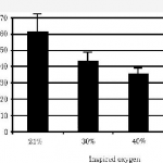
Since this topic just won’t seem to die, I’m going to try to kill the last papers I’m aware of on this topic today and tomorrow. Today’s was published in a pediatric surgical journal (!), and it’s another rabbit study. This one adds a wrinkle to the one I discussed yesterday. Not only did they inject air to create a pneumothorax (20cc this time), they punctured the pleura with a needle to create an air leak to simulate a real clinical problem.

They saw the same trend as posted yesterday, although the times were longer. Once again, resolution was measured with chest xray (performed every 12 hours this time). Unfortunately, 7 of the 27 rabbits used in each group died, leaving only 6 or 7 in each of 3 groups for analysis (room air, 40%, 60% O2). Even with wide standard deviations, the authors claimed significant differences in recovery.

Same problems as yesterday, particularly with how resolution of pneumothorax is determined. And don’t use rabbits! A bigger issue is that this is not really a clinically relevant model. First, creating an air leak would defeat the overall purpose of giving high O2 concentrations. If 60% O2 leaked into the pleural space, there would be less nitrogen to wash out so one would think that resolution would take longer. And no one would consider treating a patient with an air leak without some type of drainage device for fear of a tension pneumothorax.

Bottom line: Still not enough evidence to support this seemingly benign treatment. Tomorrow I’ll look at the (hopefully) last paper on the topic since the beginning of time, published in 1971.

Related posts:

Reference: Supplemental oxygen improves resolution of injury-induced pneumothorax. J Pediatric Surg 35(6):998-1001, 2000.

Thanks to Jonathan St. George for his comments on yesterday’s post!

Treating Pneumothorax With Oxygen (Again)

The topic of treating pneumothorax with high inspired oxygen concentrations keeps coming up! I’ve written about this a few times in the past, and the literature I found supporting the practice was terrible. Some readers brought three more studies to my attention that support it, so I’m going to take the next three days to see if there is any hope for this practice.

Today’s paper used a rabbit model where each animal was given a complete pneumothorax by the injection of 15cc (!!) of air into one hemithorax. The authors then let the pneumothorax resolve using room air or 30%, 40%, or 50% FIO2. Each group consisted of 10 rabbits, and repeat chest xrays were obtained every 6-8 hours to follow resolution.

The statistical analysis was interesting and unusual. Because the authors were studying the time to resolved pneumothorax with higher inspired O2, they were looking for a test that would analyze an “ordered alternative.” The Jonckheere-Terpstra test was used, which I have never heard of, but I’ll assume it’s the legitimate one to use.

The figure at the top of this post shows the results. Looks promising right? There was a big improvement from room air to 30%, but lesser improvement using higher oxygen concentrations. The error bars (standard error of the mean) are remarkably tight, but this makes sense since xrays were only being taken every 6-8 hours.

The two big problems with this study are that: 1. they’re rabbits and it only takes 15cc of air to drop the entire lung, and 2. standard xray is being used to measure resolution. Trying to pick apart the exact time to resolution of a 15cc pneumothorax is very difficult, and to try to do it with a test that we know is not great at detecting small amounts of air even in big humans just doesn’t work. 

Bottom line: Fancy statistics and nice looking results don’t make up for an animal model that doesn’t necessarily correlate with humans and deriving results using an inaccurate diagnostic test. Tomorrow, I’ll look at a paper in the Journal of Pediatric Surgery to see if it fares any better.

Related posts:

Reference: Resolution of experimental pneumothorax in rabbits by graded oxygen therapy. J Trauma 45(2):333-334, 1998.

Thanks to Stephanie Taft MD at Regions Hospital for finding these fine studies for me.

Best Of: High Inspired O2 Is Not An Effective Pneumothorax Treatment

The use of high concentrations of inspired oxygen seems to be a time-honored technique for trying to avoid chest tube insertion for pneumothorax. But does it stand up to scrutiny, or is this just an urban legend?

This recommendation is based upon a single case report involving 8 patients in 1983! Six patients with a pneumothorax of less than 30% showed a decrease in size of 4.2% per day on average. The two patients with pneumothoraces larger than 30% did not respond. A response was only seen with oxygen administered by a partial nonrebreather mask, not by nasal cannula.

What’s the problem? First, this is a very small case report. There were no controls, so it is entirely possible that the resolution rate without treatment was the same as that seen in this report. Furthermore, this study was performed prior to the availability of chest CT. Therefore, the true size of the pneumothoraces is only a guess since volumetric calculations could not be performed. It is not possible to distinguish a 4% change in the size of a pneumothorax by regular chest xray (click here for more details).

The bottom line: If the patient needs supplemental oxygen for management of other pulmonary conditions, then administer it. It is not indicated as an independent treatment for pneumothorax, and its use for this condition should be abandoned!

Related posts:

Reference: Noninvasive treatment of pneumothorax with oxygen inhalation. Chadha TS. Respiration 44(2):147-52, 1983

Best Of: What Percent Pneumothorax Is It?

Frequently, radiologists and trauma professionals are coerced into describing the size of a pneumothorax seen on chest xray in percentage terms. They may something like “the patient has a 30% pneumothorax.”

The truth is that one cannot estimate a 3D volume based on a 2D study like a conventional chest xray. Everyone has seen the patient who has no or a minimal pneumothorax on a supine chest xray, only to discover one of significant size with CT scan.

Very few centers have the software that can determine the percentage of chest volume taken up with air. There are only two percentages that can be determined by viewing a regular chest xray: 0% and 100%. Obviously, 0% means no visible pneumothorax, and 100% means complete collapse. Even 100% doesn’t really look like 100% because the completely collapsed lung takes up some space. See the xray at the top for a 100% pneumothorax.

If you line up 10 trauma professionals and show them a chest xray with a pneumothorax, you will get 10 different estimates of their size. And there aren’t any guidelines as to what size demands chest tube insertion and what size can be watched.

The solution is to be as quantitative as possible. Describe the pneumothorax in terms of the maximum distance the edge of the lung is from the inside of the chest wall, and which intercostal space the pneumothorax extends to. So instead of saying “the patient has a 25% pneumo,” say “the pneumothorax is 1 cm wide and extends from the apex to the fifth intercostal space on an upright film.”

Managing Chest Tube Air Leaks

There are a lot of opinions and not so much literature on how to manage chest tube air leaks. Here is some practical advice on how to deal with this occasional problem.

Most air leaks are an alveolar-pleural fistula, representing a connection between a very small airspace and the pleura. This should not be confused with a bronchopleural fistula, which involves larger airways and is much more challenging to manage.

First, identify what kind of leak it is. Remember, dry seal chest tube systems will not show an air leak unless it has a fluid chamber that can be filled with water (see related post below).

  • Expiratory – occurs during normal expiration only
  • Forced expiratory – occurs only with coughing
  • Inspiratory – occurs during inspiration in ventilated patients
  • Continuous

Inspiratory leaks are rare and should be managed conservatively with maneuvers to minimize airway pressures. Continuous air leaks can be monitored, but may indicate a bronchopleural fistula.

Expiratory and forced expiratory types account for about 98% of all air leaks. Small air leaks should be managed with water seal, not with increased suction. The main concept is to reduce air flow through the fistula so it can heal. A prospective study has shown that this technique stops small to moderate size leaks sooner than leaving on suction.

Larger air leaks will probably not seal on their own and are probably not safe to place on water seal. They will likely require pleurodesis, either chemical or mechanical via a VATS procedure. Blood and fibrin patches have also been tried.

Any air leak that extends hospital stay should be evaluated for appropriateness of discharge with a Heimlich valve or VATS pleurodesis.

References:

  • A prospective algorithm for the management of air leaks after pulmonary resection. Ann Thoracic Surg 66:1726-1731, 1998.
  • Prospective randomized trial compares suction versus water seal for air leaks. Ann Thoracic Surg 71:1613-1617, 2001.

Related posts: