Tag Archives: pneumothorax

What Percent Pneumothorax Is It?

What percent pneumothorax?

Frequently, radiologists and trauma professionals are coerced into describing the size of a pneumothorax seen on chest xray in percentage terms. They may say something like “the patient has a 30% pneumothorax.”

The truth is that one cannot estimate a 3D volume based on a 2D study like a conventional chest xray. Everyone has seen the patient who has no or a minimal pneumothorax on a supine chest xray, only to discover one of significant size with CT scan.

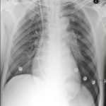
Very few centers have or use the software that can determine the percentage of chest volume taken up with air. There are only two percentages that can be determined by viewing a regular chest xray: 0% and 100%. Obviously, 0% means no visible pneumothorax, and 100% means complete collapse. Even 100% doesn’t really look like 100% because the completely collapsed lung takes up some space. See the xray at the top for a 100% pneumothorax.

If you line up 10 trauma professionals and show them a chest xray with a pneumothorax, you will get 10 different estimates of their size. And there aren’t any guidelines as to what size demands chest tube insertion and what size can be watched.

Bottom line: The solution is to be as quantitative as possible. Describe the pneumothorax in terms of the maximum distance the edge of the lung is from the inside of the chest wall, and which intercostal space the pneumothorax extends to. So instead of saying “the patient has a 25% pneumo,” say “the pneumothorax is 1 cm wide and extends from the apex to the fifth intercostal space on an upright film.”

Not Your Usual Pneumothorax?

You’ve been called to the ED to see a patient with a “spontaneous pneumothorax”, but once you meet him you see that he doesn’t fit the classic profile (tall, slim male). What gives?

After closer questioning, he admits to have been smoking crack cocaine at the time. Freak coincidence?

There are a number of case reports dating from 1984 describing this association. A number of reasons have been cited:

  • A high incidence of tobacco smoking
  • Bullous disease caused by inhaled drug use
  • Inhalation of hot gas followed by frequent Valsalva maneuvers

I’ve seen this presentation about 5 times in my career. I always ask about drug use so I can ensure that a chemical dependency screen is ordered.

Reference: Pneumothorax, pneumomediastinum, and pneumopericardium following Valsalva’s maneuver during marijuana smoking. N Y State J Med 84(12):619-20, 1984.

The Deep Sulcus Sign

Pneumothorax is frequently difficult to diagnose in the resuscitation room. Sometimes it is obvious, with a hypoxic patient and absent breath sounds. But not usually. Most of the time we rely on a chest xray to help make the diagnosis.

Unfortunately, the good old chest xray only shows a pneumothorax about 30-50% of the time. A big part of the problem is that our patients are usually supine to protect their spine. A small pneumothorax make float anteriorly in the supine position, and if it is not big enough to wrap around the lateral edge of the lung, it may remain invisible. So you need to look for gross and subtle signs on the image that will help make the diagnosis. The deep sulcus sign is one of the more subtle signs. 

Simply stated, the deep sulcus sign is a radiolucent (dark) lateral sulcus where the chest wall meets the diaphragm. The amount of lung in this area is less, so a small amount of air will tend to darken the area making it more prominent. Look at patient left in the left photo, and compare to their right side. It is much darker and appears to extend lower than usual. In more extreme cases, the amount of air just above the diaphragm may make it appear inverted (right photo).

Bottom line: If you see a deep sulcus sign on the chest xray image, strongly consider pneumothorax. If the patient begins to have hemodynamic problems, needle the chest and chase with a chest tube. If they remain stable, the patient will still require a chest tube. Chest xray always underestimates the true size of the pneumothorax. Place the usual size chest tube and manage per your usual protocol. And, as always, use your best sterile technique and definitively identify the proper side before placing the tube.

Related posts:

Best Of: Flying After Pneumothorax

Patients who have sustained a traumatic pneumothorax occasionally ask how soon they can fly in an airplane after they are discharged. What’s the right answer?

The basic problem has to do with Boyle’s Law (remember that from high school?). The volume of a gas varies inversely with the barometric pressure. So the lower the pressure, the larger a volume of gas becomes. Most of us hang out pretty close to sea level, so this is not an issue.

However, flying in a commercial airliner is different. Even though the aircraft may cruise at 30,000+ feet, the inside of the cabin remains considerably lower though not at sea level. Typically, the cabin altitude goes up to about 8,000 to 9,000 feet. Using Boyle’s law, any volume of gas (say, a pneumothorax in your chest), will increase by about a third on a commercial flight. 

The physiologic effect of this increase depends upon the patient. If they are young and fit, they may never know anything is happening. But if they are elderly and/or have a limited pulmonary reserve, it may compromise enough lung function to make them symptomatic.

Commercial guidelines for travel after pneumothorax range from 2-6 weeks. The Aerospace Medical Association published guidelines that state that 2-3 weeks is acceptable. The Orlando Regional Medical Center reviewed the literature and devised a practice guideline that has a single Level 2 recommendation that commercial air travel is safe 2 weeks after resolution of the pneumothorax, and that a chest xray should be obtained immediately prior to travel to confirm resolution.

Bottom line: Patients can safely travel on commercial aircraft 2 weeks after resolution of pneumothorax. Ideally, a chest xray should be obtained shortly before travel to confirm that it is gone. Helicopter travel is okay at any time, since they typically fly at 1,500 feet or less.

References:

  • Practice Guideline, Orlando Regional Medical Center. Air travel following traumatic pneumothorax. October 2009.
  • Medical Guidelines for Airline Travel, 2nd edition. Aerospace Medical Association. Aviation, Space, and Environmental Medicine 74(5) Section II Supplement, May 2003.

Pneumothorax And Oxygen: The Final Post!

Okay, this is the last time I’m going to write about this. Hopefully I can provide the final nail in the coffin for this idea. Previously, the oldest paper I could find that was cited as a reason to use high inspired oxygen to treat pneumothorax was from 1983. I found what I think is the earliest (and the last that I will discuss) from 1971!

Twelve patients were retrospectively reviewed who recovered without intervention from a spontaneous pneumothorax. Another 10 were monitored prospectively with the same condition, but were given “high concentration oxygen” (??) by mask from 9 to 38 hours at a time. During intervening periods, the patients breathed room air. Daily chest xrays were obtained, and here is the cool part:

The inner edge of the chest wall and the outer edge of the lung were traced on transparent paper. This was then superimposed on graduated graph paper and the area corresponding to the pneumothorax cavity was measured. The rate of absorption was expressed in cm2/24 hrs.

Need I say more? The authors did show graphically that the apparent rate of absorption tripled in the treated patients, from about 5cm2/day to about 15cm2/day, and was higher in patients with a larger pneumothorax. The problem here is the same as before: chest xray does not allow volumetric estimates, so any results relying on them are suspect. At least it’s not a rabbit study.

Bottom line: There’s just no convincing data to support this practice, so let’s stop using it. Simple physics suggests that this should work, but the effect is just not clinically significant enough to offset the possibility of mishaps from an inpatient admission for oxygen therapy (see yesterday’s post). As I mentioned yesterday, look at the clinical status of your patient. If they have any detectable blood in their chest, they’ll probably need drainage. If not, and if they feel normal, discharge and follow up with a repeat xray in a week. The pneumo will probably be gone. If they do have some compromise, then insert the smallest tube you can. If done properly and a one-way valve can be used, the patient may still be managed as an outpatient.

Related posts:

Reference: Oxygen therapy for spontaneous pneumothorax. Br Med J 4:86-88, 9 Oct 1971.