Tag Archives: pediatric

ED Thoracotomy: Kids ARE Just Small Adults

You’ve undoubtedly read this trite phrase somewhere in your training: “Kids aren’t just small adults!” There are many examples where this is absolutely true. Think about arterial extravasation in solid organ injury. Or severe traumatic brain injury. There are major differences in treatment aggressiveness for both of these.

But what about the code situation? I’ve noted a peculiar phenomenon over the years with regard to pediatric codes of all kinds. Adults tend to persist far longer at resuscitative efforts over children than they normally would on other adults. And what about that most extreme code situation, the emergency thoracotomy?

I’ve also seen the use of this procedure in children who don’t meet the usual adult criteria. But they are kids, right? They can bounce back from more severe insults, right? I hope that I’ve convinced you over the years that one can’t just assume and generalize anything. Things that seem like so much common sense often turn out to be wrong. Think back to the days of the stress / spicy food theory of peptic ulcer disease. This seems so silly now that we recognize the role of H. Pylorii.

Scripps Mercy adult and Rady Children’s Hospital pediatric trauma centers in San Diego performed an extensive review of the National Trauma Data Bank over a three year period. They focused on patients 16 years of age or less who underwent ED thoracotomy within 30 minutes of arrival at the trauma center. They focused on procedure indications and the eventual outcomes.

Here are the factoids:

  • A total of 114 patients were recorded in the NTDB, with a mean age of 10 years and median Injury Severity Score of 26 (this is the three year experience in the entire US in three years!)
  • Males were disproportionately involved at 69%, although this is less than in adults
  • Thoracotomy was performed promptly, with a median time after arrival of 5 minutes
  • Mechanism of injury was almost evenly split between penetrating (56%) and blunt (44%)
  • Blunt mechanism mortality was 94% vs 88% for penetrating
  • Penetrating injury outside of the thorax was uniformly fatal
  • Patients without signs of life on arrival, regardless of mechanism, also had a 100% mortality rate
  • Treatment at an adult trauma center, freestanding pediatric center, or combined center had no impact on these dismal outcomes

Bottom line: This is an interesting paper, and shows that the outcomes after ED thoracotomy in kids is even more dismal than in adults. This is particularly true for children arriving without vital signs and for penetrating abdominal trauma.

However, the authors go on to suggest a practice guideline for pediatric emergency thoracotomy similar to the EAST adult guidelines based on their study findings. However, I think this is ill advised. Have a look at the absolute numbers:

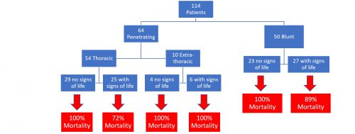
The largest subgroup has only 29 patients in it. These numbers are way too small to consider a guidelines change.

This paper shows that kids are just small adults when it comes to ED thoracotomy. And they seem to do even more poorly with no vital signs or penetrating injuries outside of the chest. So think carefully the next time you must consider this procedure in a child.

Reference: Nationwide Analysis of Resuscitative Thoracotomy in Pediatric Trauma Time to Differentiate from Adult Guidelines? J Trauma published ahead of print, July 6, 2020.


Mainstem Intubation In Pediatric Patients: How To Avoid It

In my last post, I reviewed a simple technique utilizing a reminder card and provider feedback loops to reduce deep intubations in pediatric patients. Today, I’ll review three other techniques and discuss a paper that compared their efficacy.

A variety of techniques for determining and/or confirming endotracheal tube position exist. Use of one or more of them is important in children due to their short trachea and increased likelihood of deep intubation. Some, like the confirmatory chest x-ray, are obvious. However, it’s more desirable to apply techniques during the intubation in order to avoid deep intubation in the first place. Hyperinflation of one lung, especially in very small children, can cause a host of impairments and complications that may compound their other injuries.

A paper from the University Hospital Basel in Switzerland evaluated three techniques: bronchoscopic insertion to a specific depth, cuff palpation in the sternal notch, and intentional right mainstem intubation followed by slow withdrawal during auscultation.  Each of 68 children ranging in age from 0 to 4 years were studied using all three techniques.

Each endotracheal tube was marked at the ideal insertion point that would ideally be placed just beyond the vocal cords. The distance from this mark to the mouth end of the tube was measured so actual intubation depths could be compared.

Bronchoscopic insertion was always performed first to obtain a baseline depth measurement, essentially the gold standard. The other two techniques were performed in random order. For the cuff palpation technique, the trachea was palpated while the balloon was intermittently partially inflated until it could be felt at the suprasternal notch. For the mainstem intubation technique, the uninflated tube was advanced until breath sounds in the left axilla disappeared. It was then slowly withdrawn until sounds reappeared.

Distances from the tip of the tube to the carina was calculated using the insertion depth at the incisors and the initial ideal intubation depth mark. Here are the factoids:

  • Insertion to a depth mark on the tube via bronchoscope technique resulted in the highest tube tip with respect to the carina, and also with the greatest depth variability
  • The cuff palpation technique resulted in less distance to the carina (about 19mm vs 36 for the mark technique) and less variability
  • Use of the mainstem intubation with pullback technique resulted in the tube tip resting within just a few mm of the carina, but tube depth was very consistent

Bottom line: What to make of all this? Which technique is “best?” First, it’s not practical or advisable to use a bronchoscope for every pediatric intubation. It’s invasive and adds complexity and time to a critical procedure. The cuff palpation technique also takes additional time due to the repeated cuff inflation/deflation that is required. However, the tube position is fairly accurate and safe.

The intentional right mainstem intubation with pull-back seems a bit sketchy. It requires some type of ongoing ventilation while the tube is being inserted, as well as someone who can listen to the left chest. Additionally, it results in a tube position that is so low that neck positioning may move it into the mainstem bronchus again.

In my mind, estimation of the proper depth pre-intubation is probably the best. Strict attention must be paid to the final depth of the tube once it is inserted, as measured by the distance marker at the incisors. This number must match the one decided upon at the start of the procedure. A good exam of the chest should be carried out to quickly identify an inadvertent mainstem intubation. And finally, a quick confirmatory chest x-ray should always be obtained for objective information on tube position. 

The fancy techniques described in this paper add too much time and complexity for intubation in a trauma situation. They may very well have a place in the OR where the situation is more controlled and there is more advanced equipment and support. But stick to the basics when intubating children in your trauma bay!

Reference: Assessment of three placement techniques for individualized positioning of the tip of the tracheal tube in children under the age of 4 years. Ped Anesthesia 25:379-285, 2014.

Mainstem Intubation In Pediatric Patients: How Common?

Mainstem intubation in the pediatric patient is a common problem. There are two major issues: the trachea is shorter than in an adult, and the angles are different making intubation of the right mainstem bronchus much easier. Frequently, the intubator watches the balloon slide between the cords, then pushes the tube in “just a little further.”

Unfortunately, that “little bit” can vary significantly. An abstract from my hospital was presented at the Pediatric Trauma Society in 2016. Subjectively, we noticed that mainstem intubation was occurring with some regularity in our pediatric trauma patients.  It seemed as though insufficient attention was being paid to the depth of the tube.

A major difference between adult and pediatric intubations is that in adults, optimal tube depth is locked into a relatively narrow range. In children, the depth varies considerably based upon child age and size. And small variances in depth can have major implications for tube position.

We decided to implement a PI project to change our intubation policy. In order to focus the entire team on tube depth, a color coded card was attached to each size of endotracheal tube. This card listed the optimal depth for insertion. Once the provider inserted the tube, the final depth was called out for the team and documentation scribe to hear. This had the added advantage of allowing multiple team members confirm the appropriateness of tube depth. A chest x-ray was immediately obtained to confirm position.

We retrospectively reviewed our seven year experience with pediatric intubations, from 2009-2015. Here are the factoids:

  • Nearly 2,000 pediatric trauma patients were admitted during the study period
  • 94 patients (5%) required intubation in the ED
  • Prior to implementation of the new protocol, 6 of 68 patients (8.8%) had confirmed right mainstem intubation 
  • After the change, only one further mainstem intubation occurred in 26 procedures (3.8%)

Bottom line: Unfortunately, this series is too small to determine statistical significance. There is a definite trend toward fewer mainstem intubations. It appears that by calling more attention to the proper tube depth, fewer deep placements occur. Our numbers  have remained low since this change.

Are there other methods to ensure proper ET tube placement in small patients? In my next post, I’ll review a paper that compares three additional different techniques that can be used.

Reference: Eliminating the Preventable Occurrence of Right Mainstem Intubation in the Pediatric Trauma Patient: A Quality Performance Improvement (PI) Initiative. Pediatric Trauma Society Poster Abstract #1, 2016.

AAST 2019 #3: Delayed Splenectomy In Pediatric Splenic Injury

Nonoperative management of the blunt injured spleen is now routine in patients who are hemodynamically and have no evidence of other significant intra-abdominal injury.  The trauma group at the University of Arizona – Tucson scrutinized the failure rate of this procedure in children because it is not yet well established.

They reviewed 5 years of data from the National Readmission Database. This is actually a collection of software and databases maintained by the federal government that seeks to provide information on a difficult to track patient group: those readmitted to hospitals after their initial event.

Patients who had sustained an isolated spleen injury who were less than 18 years old and who had either nonoperative management (NOM), angioembolization (AE), or splenectomy were analyzed. Outcome measures included readmission rate, blood transfusion, and delayed splenectomy. Common statistical techniques were used to analyze the data.

Here are the factoids:

  • About 9500 patients were included, with an average age of 14
  • Most (77%) underwent NOM, 16% had splenectomy, and 7% had AE (no combo therapies?)
  • Significantly more patients with high grade injury (4-5) had splenectomy or AE than did the NOM patients (as would be expected)
  • A total of 6% of patients were readmitted within 6 months of their initial injury: 12% of NOM *, 8% of AE *, and 5% of those with splenectomy (* = statistically significant)
  • The NOM and AE patients were also more likely to receive blood transfusions during their first admission
  • Delayed splenectomy occurred in 15% of cases (7% NOM and 5% AE) (these numbers don’t add up, see below)
  • Statistical analysis showed that delayed splenectomy was predicted by high grade injury (of course), blood transfusion (yes), and nonoperative management (huh?)
  • In patients who were readmitted and splenectomized, it occurred after an average of 14 days for the NOM group and 58 days for AE (huh?)

The authors concluded that “one in seven children had failure of conservative management and underwent delayed splenectomy within 6 months of discharge.” They stated that NOM and AE demonstrated only a temporary benefit and that we need to be better about selecting patients for nonoperative management.

Hmm, there are several loose ends here. First, what is the quality of the study group? Was it possible to determine if these patients had been treated in a trauma center? A pediatric vs adult trauma center? We know that there are outcome disparities in spleen trauma care at different types of trauma centers. 

Next, are they really pediatric patients? Probably not, since age < 18 were included and the average age was 14. Injured spleens in pre-pubescent children behave much better than adolescents, which are more adult-like.

And what about the inherent bias in the “readmission data set?” You are looking only at patients who were readmitted! By definition, youare looking at a dataset of poorer outcomes. What if you had identified 9,500 initial patient admissions from trauma registries and then tried to find them in the readmission set. I know it’s not possible to do that, but if it were I would bet the readmission and delayed splenectomy numbers would be far, far lower.

And what about those delayed splenectomy numbers? I can’t get the percentages to match up. If 15% of the 7965 patients who didn’t have an initial splenectomy  had it done later, how does 7.2% of the 7318 NOM patients and 5.3% of the 1541 AE patients add up?

Bottom line: The usual success rate tossed around for well-selected nonoperative management is around 93% when optional adjunctive AE is part of the algorithm. That’s a 1 in 14 failure rate, and it generally occurs during the initial hospitalization. In my experience, readmissions are very rare. And that’s for adults; children tend to behave even better!

I wouldn’t consider changing my practice yet based on these findings, but the devil will probably be in the details!

Here are some questions for the presenter and authors:

  • Please provide some detail on the data set. We really need to know an age breakdown and the types of centers they were treated at, if available.
  • Discuss the potential data set bias working backwards from a database that includes only readmitted patients.
  • Please clarify the delayed splenectomy statistics to help match up the numbers.

I’m anticipating a great presentation at the meeting!

Reference: Delayed splenectomy in pediatric splenic injuries: is conservative management overused? AAST 2019 Oral abstract #8.

Button Batteries: Part 2 – Getting Them Out

In my last post, I detailed how to suspect and image a button battery ingestion. In this one, I’ll describe how to extract them, and how quickly it’s necessary.

When batteries come to rest and are surrounded by moist mucosal tissue, a current arc is generated around the two sides of the button. This releases heat, which coagulates the surrounding tissue. Depending on the location, closeness of contact, and the duration, these burn injuries may extend into underlying tissue. This is of particular significance in the esophagus, which is in close proximity to the thoracic aorta.

Here’s a simple demonstration you can do at home with some lunch meat:

Here are guidelines for what to do when you encounter pediatric patients who have ingested a button battery:

  • If the child is experiencing bleeding from the upper GI tract, activate your trauma team. The child may have an aorto-esophageal fistula. If there is no active bleeding, obtain a chest x-ray to assess the battery’s position. If there is active bleeding, proceed to the OR (preferably a hybrid room if you have one) and use fluoro to locate the battery. If bleeding persists, call appropriate pediatric surgical specialists (surgery, CV surgery, GI), activate your massive transfusion protocol, and consider tamponade with a Blakemore tube (remember those?) or a urinary catheter if you don’t have one.
  • No bleeding from the upper GI tract? If the battery is large (>20mm) and/or the child is small (<5 years), and is lodged in the esophagus, proceed immediately to OR and remove endoscopically.
  • Batteries in the stomach are of less concern. They will generally pass if <20mm. A repeat x-ray after 48 hours should be obtained for larger batteries. If still in the stomach, they should be removed endoscopically. Smaller batteries will usually pass, and should be re-imaged after two weeks to confirm this.

References:

  • Button battery and magnet ingestions in the pediatric patient.  Curr Opin Pediatrics 30:653-659, 2018.
  • Management of ingested foreign bodies in children: a clinical report of the NASPGHAN Endoscopy Committee. J Pediatric Gastroenterol Nutr 60:562-574, 2015.