Tag Archives: MRI

MRI And External Fixators

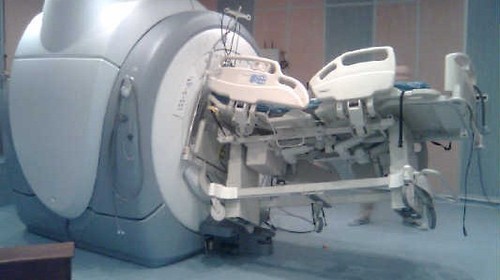
MRI is an indispensable tool for evaluation of spine and soft tissue trauma. However, a great deal of effort was be made to ensure that any patient scheduled for this test is “MRI compatible.” The fear is that any retained metallic fragments may move or heat up once the magnets are activated.

But what about trauma patients with external fixators? That is one big hunk of metal that is inserted deep into your patient. There are three major concerns:

  • Is the material ferromagnetic? If so, it will move when the magnets are activated and may cause internal injury. These days, there are many fixator sets that are not ferromagnetic, avoiding this problem.
  • Can currents be induced in the material, causing heating? This is not much of a problem for small, isolated objects. However, external fixators are configured in such a way that loops are created. The fluctuating magnetic fields can induce currents that in turn will heat the surrounding tissue. And thinner materials (narrow pins) result in more current and more heating.
  • Will the metal degrade image quality?

The biggest challenge is that there is no standard ex-fix configuration. Our orthopaedic colleagues get to unleash their creativity trying to devise the appropriate architecture to hold bones together so they can heal properly. This makes it difficult to develop standardized guidelines regarding what can and can’t go into the scanner.

However, there is a growing body of literature showing that the heating effects are relatively small, and get smaller as the distance from the magnet increases. And non-ferromagnetic materials move very little, if at all, and do not interfere with the image. So as long as nonferromagnetic materials are used, the patients are probably safe as long as basic principles are adhered to:

  • Other diagnostic options should be exhausted prior to using MRI.
  • Informed consent must be obtained, explaining that the potential risks are not completely understood.
  • The fixator must be tested with a handheld magnet so that all ferromagnetic components can be identified and removed.
  • All traction bows must be removed.
  • Ice bags are placed at all skin-pin interfaces.
  • The external fixator must remain at least 7cm outside the bore at all times.

Bottom line: MRI of patients with external fixators can be safely accomplished. Consult your radiologists and physicists to develop a policy that is specific to the scanners used at your hospital. 

Related posts:

Best of EAST #3: Spine MRI Usage After EAST Guidelines

In 2015, EAST published their practice guidelines for spine clearance in the obtunded blunt trauma patient. Click here to view them. They stated that a high-quality CT scan can be used to remove (clear) the cervical collar in these patients. This avoids the use of the expensive and personnel-intensive MRI clearance.

The group at UCSF used the NTDB to review the use of MRI in such patients over an 11 year period. They focused on comatose patients (GCS < 8) with an AIS head > 3 and intubation for more than 72 hours. They used logistic regression to equalize confounders while examining the use of MRI over time, before and after the guidelines were published.

Here are the factoids:

  • More than 75,000 patients from 530 trauma centers were included
  • Patients who were older, Hispanic, uninsured, or involved in a car crash were less likely to undergo spinal MRI
  • Level I centers were more likely to use MRI for clearance than Level II centers
  • Patients evaluated after release of the practice guidelines were 1.7x more likely to undergo MRI for spine clearance (!!)

The authors concluded that spinal MRI use has been increasing since 2007 despite publication of the EAST guideline.

My comments: To me, this indicates one of the following:

  1. Nobody reads the EAST guidelines, or
  2. Trauma programs believe that they alone are able to figure out what is right, and everyone else is wrong

I suspect that it is #2. For some reason, trauma programs insist on doing it their own way despite what decent evidence shows. I think that this represents a defense mechanism to minimize the cognitive dissonance that comes with defying what is published in the literature.

I always encourage programs to borrow/steal what is already out there when crafting their own practice guidelines. Someone else has already done the work, why not take advantage of it? Typically, it’s just an excuse to continue doing things the way they’ve always been done.

This incessant reinventing the wheel becomes tiresome. And for once, I don’t have many questions or suggestions for the authors. Their evidence is pretty well laid out. 

My questions for the author / presenter are:

  1. Do you use MRI for spine clearance in your obtunded blunt trauma patients? And if so, WHY?
  2. Why do you think there are demographic and trauma center level disparities? Is it the teaching environment? Something else?

To everyone else, I say “get over yourself and read the literature!”

Reference: Assessing the e3ffect of the EAST guideline on utilization of spinal MRI in the obtunded adult blunt trauma patient over time. EAST 2021, Paper 7.

What? Still Using MRI For Cervical Spine Clearance?

Cervical spine clearance as evolved considerably over the years. First, there were five views of the spine using plain radiography. Then there were three. Then we moved to CT scan with clinical clearance. And currently, many institutions are relying only on CT.

But MRI has been used as an adjunct for quite some time. Initially, it was the tie breaker in patients who had equivocal CT findings, and for a while it was used for clearance in obtunded patients. And thanks to conflicting literature and disparate studies, the occasional usage became more frequent.

The group at Cedars-Sinai Medical Center in Los Angeles  noted that the percentage of patients undergoing MRI for cervical spine evaluation at their center slowly slowly crept up from 0.9% to 5.6% over a 10 year period. They designed a study to analyze the utility of this practice and inform their future practice.

Here are the factoids:

  • Over 9,000 patients had cervical spine CT during the 10-year study period; 513 (5.6%) were positive
  • Of the 513 CT-positive patients, 290 (56%) underwent an MRI. This showed:
    • Confirmation of the major injury in 250
    • Minor injury in 40
    • Clinically significant injury was seen in only 2 which was no surprise since they both had neurologic deficits
  • Of the 8,588 CT-negative patients, only 9 had clinically significant findings and 8 of them had neurologic deficits

Bottom line: So what have we learned here? First, MRI usage at Cedars-Sinai increased over time but was really not that useful. The main use was for imaging obtunded patients or those with an obvious neurologic deficit.

More than half of patients with positive CT scans also underwent MRI. If a major injury was seen on CT, MRI confirmed it. But if the CT findings were minor, none of the MRIs added any clinically significant findings in the absence of a neurologic deficit.

And what about MRI after negative CT? In the absence of a deficit, only one had a clinically significant finding (which only required a brace).

This study shows the wisdom of monitoring “how we do it.” There is sometimes some creepage away from what the literature shows is the best practice. The best way to remedy this is to do a good study, just like the authors did. They saw a slow change in practice, investigated it, and found that there was no good clinical reason for it. This gives the trauma program the ammunition to squelch the unwelcome behavior and return the clinicians to best practices.

Reference: Is MRI becoming the new CT for cervical spine clearance? Trends in MRI utilization at a Level I trauma center. J Tra publish ahead of print, DOI: 10.1097/TA.0000000000002752, 2020.

Are You Still Using MRI To Clear The Cervical Spine?

There is a fairly robust  amount of data that shows that, properly performed, the cervical spine can be cleared using a high quality CT read by a highly skilled radiologist. This is true even for obtunded patients. Pooled data suggest that the miss rate in this group is only 0.017%. And MRI is not perfect either, missing significant ligamentous injury in a small number of patients.

But it seems that some trauma professionals are still using MRI in some cases despite this data. The latest study on MRI focuses on the cost-effectiveness of the technique. The authors selected patients with GCS < 13 to be their obtunded group, which is probably a bit high. Nevertheless, they used a fairly sophisticated (meaning hard to understand) modeling-based decision analysis using a computerized simulation. This allowed them to compare different clearance strategies without performing large randomized clinical trials.

The authors considered MRI vs no MRI, false results, collar use and complications, MRI use with cost and complications, and the worst-case scenario of tetraplegia. Here is a flow chart of the scenarios considered. (Courtesy JAMA Surgery)

Here are the factoids:

  • The mean cost for followup vs no followup was $14K vs $1K, with no increase in quality adjusted life years (QALY)
  • No followup was the better strategy when the negative predictive value of CT was high (>98%), when the risk of an unstable injury treated with a collar turning into a permanent deficit was >25%, or if the chance of a missed injury becoming a permanent deficit was >58%
  • No followup MRI was the better strategy in all 10,000 iterations of the simulation

Bottom line: Yes, this is a fairly heavy computer simulation. But the reality is that we will never be able to design a large enough study to critically evaluate this issue and have it pass any IRB review. So it’s probably as good as it will ever get. It’s time to stop wasting money and putting obtunded patients in harm’s way by locking them into a relatively inaccessible MRI scanner for 30 minutes just to confirm the CT. Or keeping a collar until until the skin breaks down.

Here is a copy of the practice guideline we use for clearing all cervical spines, obtunded or not. Yes, there is some weirdness with soft collars, which mainly serve as a reminder to re-examine the patient at some point. But note the scan technique and requirement that it be read by a neuroradiologist for final clearance.

Related link:

Reference: Cost-effectiveness of Magnetic Resonance Imaging in Cervical
Clearance of Obtunded Blunt Trauma After a Normal
Computed Tomographic Finding

Using MRI To Predict Outcome From Diffuse Axonal Injury (DAI)

Has this happened to you? A patient with a serious head injury is not waking up as expected. There were a few punctate hemorrhages seen on the initial CT scan. Your neurosurgery colleague orders an MRI to “provide a prognosis on the patient’s injury.”

Is this a legitimate request? Sure, MRI is very sensitive at detecting very small hemorrhages that may signal the presence of diffuse axonal injury (DAI). But do more abnormalities on MRI equal a poorer prognosis or longer recovery time?

A group from Vanderbilt presented their data from a retrospective cohort study at EAST earlier this year.  They reviewed 7 years of data from 2006 to 2012, including all patients with a head CT positive for intracranial injury and an MRI within 2 weeks. They excluded penetrating injuries and patients with psychiatric or neurologic disorders. They analyzed information on three year mortality, functional outcome, and quality of life.

Here are the factoids:

  • A total of 311 patients met all inclusion/exclusion criteria, with a median age of 40 and serious injury (average ISS 29, average ICU length of stay 6 days)
  • Functional status at discharge could be assessed in 240 patients, and only 118 could be contacted for long-term followup questions
  • Only 56% of patients with severe TBI had an MRI positive for DAI
  • Functional status was lower on discharge for patients positive for DAI on MRI
  • There was no difference in Glasgow Outcome Score, quality of life, or 3 year survival in patients with MRI evidence of DAI compared to those without

Bottom line: This is a relatively large study, but there are still several weaknesses that could skew the numbers a bit. However, it appears that MRI for prognostication of outcomes in patients with clinical DAI is not very helpful. First, only about half with a clinical picture of DAI showed it on MRI. And sure, MRI may tell us a little bit about their status when they are discharged from the hospital to rehab or transitional care. But is that information very useful? It certainly does not help predict their outcome in the longer term. So why order an expensive and difficult study (think restraints, sedation, lots of pumps and monitors) to tell us what we already know based on our experience with severe TBI?

Reference: Prognosis of diffuse axonal injury with traumatic brain injury. J Trauma 85(1):155-159, 2018