Tag Archives: management

Best Of AAST 2022 #9: Management Of Low Grade Blunt Thoracic Aortic Injury

It has been interesting to watch the evolution of the treatment of blunt thoracic aortic injury (BTAI). In my training, patients with an abnormal mediastinal contour on chest x-ray were whisked away to interventional radiology for a thoracic aortic angiogram. Yes, there was no such thing as a CT scan for anything but the head! Here’s a sample chest x-ray:

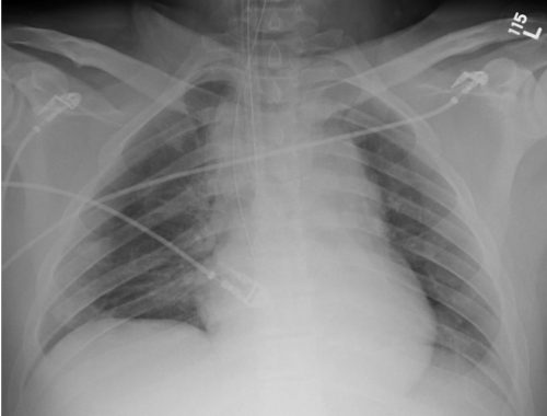
I wrote a post 12 years ago describing the various findings of a blunt aortic injury, many of which are present above. You can the post by clicking here.

In the 1-2% where the angio was positive (yes, you read that right; we did a lot of negative angiograms) the patient was then whisked off to the OR for an open aortic graft via thoracotomy. The advent of CT scans of the torso and improving resolutions allowed us to be more selective with angiography. And finally as CT angiography matured, aortic angiography became a thing of the past.

Then came TEVAR about 20 years ago. There were some growing pains as we refined the technology, but now this endovascular procedure is the standard of care for most aortic injuries. While there was also a place for nonoperative management, it was really just maintaining a reasonably low blood pressure to protect the vessel while getting the patient into good enough condition to tolerate anoperation.

Now we are beginning to slice and dice treatment based on grade. Many centers have recognized that an intimal injury (Grade I) is only a very minor disruption to the inside of the vessel and does not make it more susceptible to rupture. Grade II management has been less clear. Here is a diagram of the various grades.

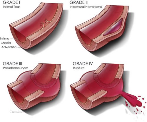
It makes sense that an invasive procedure may be less helpful for injuries that do not disrupt the layer of the vessel that provides its strength. But remember, common sense isn’t always the truth.

The group at Dell Seton Hall reviewed all patients with low grade BTAI (Grades I and II) in the Aortic Trauma Foundation Registry for a six year period. Their hypothesis was that these injuries could be successfully treated with medical management alone. They reviewed the data for mortality, complications, vent days, and lengths of stay.

Here are the factoids:

  • A total of 880 patients were enrolled and 274 had low grade injuries; 5 were then excluded when their lesion progressed and they underwent TEVAR
  • Of the 269 remaining patients, 81% were treated with medical management (81% Grade I, 19% Grade II) and the remainder with TEVAR (20% Grade I, 80% Grade II)
  • Rates of thoracotomy, craniectomy, and sternotomy were the same in both groups, but TEVAR patients were more likely to have a laparotomy (31% vs 15%)
  • Mortality was significantly higher in the TEVAR group (18% vs 8%) but the mortality from the aorta was not quite significant (4% vs 0.5%)
  • Complications (DVT and ARDS) were also significantly higher in the TEVAR group
  • Vent days and lengths of stay were equivalent

The authors concluded that medical management alone is safe and appropriate, with a lower mortality and decreased complications compared to routine TEVAR.

Bottom line: Hmm, color me skeptical. Remember, this is a registry study, so information tends to be limited outside the usual demographics and data points that are very pertinent to the purpose of the registry. The most important concept is that the patients in each group must be identical in every way except for the intervention.

We can try to make them as identical as possible by matching pairs or subgroups of patients. But the biggest problem is that the number of patient characteristics that might be important to match may not be available for analysis in the database.

When the patients were initially treated at contributing centers, there were no specific rules that the individual surgeons had to follow to decide between medical management and TEVAR. They could pick and choose based on their own experience. Could the surgeon have recognized some patients as higher risk and opted for TEVAR to make sure the aorta would not become an issue in conjunction with their other injuries? And unfortunately, perhaps that higher risk issue is what ultimately killed them and not the aorta. It’s basically a form of unhealthy user bias.

This abstract is an interesting tidbit that should push us to question whether medical management is better, at least in some subsets of low grade aortic injury patients. Then someone can perform a more robust study to confirm or refute its safety. Unfortunately, this may never happen due to the low incidence of this injury. It took six years to accumulate only 269 eligible patients in this registry!

Here are my questions for the authors and presenter:

  • What data points are actually in the registry? Specifically, is there fine detail about the other injuries the patient had? Could these have contributed to TEVAR mortality, or the selection of the patient for TEVAR to try to reduce the perceived mortality risk?
  • When you stated that there was “no difference in demographics or mechanism of injury” what were these, exactly?
  • What did patients actually die from in the Grade I and Grade II groups? Be more specific than “aortic-related” or not.
  • Do you have any worries about the five patients whose lesions progressed? When should patients be re-scanned to identify those who might benefit more from TEVAR?

This is a thought provoking abstract, and I am very interesting in hearing what the next steps should be.

Reference: MEDICAL MANAGEMENT IS THE TREATMENT OF CHOICE FOR LOW GRADE BLUNT THORACIC AORTIC INJURIES. Plenary paper #45, AAST 2022.

Practical Tip: Evaluation of Hematuria in Blunt Trauma

Bloody urine is a relatively uncommon finding in blunt trauma patients. Hematuria ranges from microscopic to gross. Microscopic means blood that can only be seen with a microscope, and gross means visible to the naked eye. In trauma, we only care about gross hematuria, which ranges from the faintest of pink to the deepest red.

In the picture above gross hematuria is present in all tubes but the far right one. Those four will need further evaluation.

In trauma, gross hematuria is a result of an injury to kidney, ureter or bladder. Blunt injury to the ureter is so rare it’s reportable, so you can pretty much forget that one unless the mechanism is extreme. So you really just need to focus on kidney and bladder.

Any victim of blunt trauma that presents with visible hematuria needs to be evaluated by CT of the abdomen and pelvis with an added CT cystogram. Standard CT technique is done without a urinary catheter, or with the catheter clamped. This is not acceptable for hematuria evaluation, as only 50% of bladder injuries show up with this technique.

CT cystogram is an add-on to the standard CT, and consists of the administration of contrast into the bladder which is then kept under pressure while the scan is performed. Delayed slices through the pelvis after the bladder is depressurized and emptied is routine. Nearly 100% of bladder injuries are detected using this technique.

If the CT shows a renal laceration or hematoma, the patient should be admitted and managed according to your solid organ injury protocol. Kidney injuries fare better that livers and spleens, and only rarely require surgery. If no kidney or bladder injury is seen, the default diagnosis of a renal contusion is the culprit. No treatment is needed, and the patient can be discharged if no other injuries are present. The blood will clear over a few days, but may disappear and reappear a few times in the process. Be sure to warn the patient that this may occur, or you may receive some surprise phone calls. The patient can followup with their primary care physician in a week or two.

The majority of these injuries do not require urologic consultation. Complex injuries with extravasation of urine out of the kidney, or injuries to the collecting system should be referred to a urologist, however.

Practical Tip: Evaluation of Hematuria in Blunt Trauma

Bloody urine is a relatively uncommon finding in blunt trauma patients. Hematuria ranges from microscopic to gross. Microscopic means blood that can only be seen with a microscope, and gross means visible to the naked eye. In trauma, we only care about gross hematuria, which ranges from the faintest of pink to the deepest red.

In the picture above gross hematuria is present in all tubes but the far right one. Those four will need further evaluation.

In trauma, gross hematuria is a result of an injury to kidney, ureter or bladder. Blunt injury to the ureter is so rare it’s reportable, so you can pretty much forget that one unless the mechanism is extreme. So you really just need to focus on kidney and bladder.

Any victim of blunt trauma that presents with visible hematuria needs to be evaluated by CT of the abdomen and pelvis with an added CT cystogram. Standard CT technique is done without a urinary catheter, or with the catheter clamped. This is not acceptable for hematuria evaluation, as only 50% of bladder injuries show up with this technique.

CT cystogram is an add-on to the standard CT, and consists of the administration of contrast into the bladder which is then kept under pressure while the scan is performed. Delayed slices through the pelvis after the bladder is depressurized and emptied is routine. Nearly 100% of bladder injuries are detected using this technique.

If the CT shows a renal laceration or hematoma, the patient should be admitted and managed according to your solid organ injury protocol. Kidney injuries fare better that livers and spleens, and only rarely require surgery. If no kidney or bladder injury is seen, the default diagnosis of a renal contusion is the culprit. No treatment is needed, and the patient can be discharged if no other injuries are present. The blood will clear over a few days, but may disappear and reappear a few times in the process. Be sure to warn the patient that this may occur, or you may receive some surprise phone calls. The patient can followup with their primary care physician in a week or two.

The majority of these injuries do not require urologic consultation. Complex injuries with extravasation of urine out of the kidney, or injuries to the collecting system should be referred to a urologist, however.

Guideline: How To Manage Bleeding In The Anticoagulated Patient

Over the past year, I’ve written about bleeding problems in trauma patients caused or exacerbated by the various anticoagulants now on the market. The field of available drugs keeps growing, and the number of ways to keep blood in liquid form is increasing.

Here’s a link to a set of guidelines for approaching and treating patients who are taking these medications and then develop problematic bleeding. There are few good studies that have actually analyzed the efficacy of these methods, but it’s what we have to work with now. 

If you have any additional maneuvers that you think should be included, please comment or email. And feel free to implement some studies to find the real best practices.

Link: Guideline for bleeding in patients taking anticoagulants

Related posts:

Rib Fracture Management

A reader sent a query yesterday regarding treatment of rib fractures, and specifically asking about epidural analgesia. Today, I’ll try to answer those questions.

Rib fractures, with or without other injuries, are a big killer in trauma patients. This is particularly true in the elderly. Overall mortality rates range from 3% to 13%, with the most import factor being pain. So what is the best way to manage patients with rib fractures to speed their safe recovery?

It’s best to attack this problem from three different directions simultaneously: pain control, respiratory hygiene (or pulmonary toilet if you’re a pessimist), and activity management.

There are many approaches to pain management, which include:

  • Oral or IV analgesics
  • Various types of blocks (intrapleural, intercostal, paravertebral, epidural)
  • Topical agents (xylocaine patch)
  • Stabilization (surgical only; belts and straps are bad for breathing)

Epidural analgesia is usually seen as the ultimate form of pain control, and is usually recommended for patients with multiple fractures or severe pain with inadequate response to medications and blocks. Much of the literature on its use is based on ICU patients who were not injured. A meta-analysis was conducted that specifically looked at epidural analgesia results in trauma patients, and found that it did improve pain management and some pulmonary function tests. However, there did not appear to be any change in mortality, ICU or hospital length of stay, or time on a ventilator.

Respiratory hygiene may involve simple measures such as coughing and deep breathing, incentive spirometry, and even mechanical ventilation in severe cases. Activity management consists of turning, sitting in a chair, walking, and forms of mechanical chest wall oscillation.

Bottom Line: The key to rib fracture management is a systematic approach that address all three dimensions of care based on objective patient measures. One size does not fit all, so more aggressive measures are warranted for more severe injury. I’ve attached an interesting patented scoring system and management algorithm, as well as two protocols from US trauma centers that range from simple (Vanderbilt) to more complex (West Virginia University).

Please feel free to comment, and I’d be happy to look at your protocol. Please email it to me!

Related post: History of epidural analgesia

Downloads

References

  • Effect of epidural analgesia in patients with traumatic rib fractures: a systematic review and meta-analysis of randomized controlled trials. Can J Anaesth 56(3):230-42, Epub 2009 Feb 11.
  • Rib Fracture Score and Protocol, US Patent #7,225,813 B2 – June 5, 2007