Tag Archives: Interesting Case

Knife To The Back! Part 2

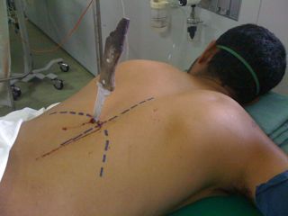
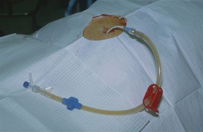
Yesterday, I presented a case of a young man with a knife in his back. He was brought to your ED in the prone position. The question was, what to do next?

With any trauma patient, regardless of size, shape, or position, the first question is always, “does this patient belong in the ED?” And usually, that question is answered by checking hemodynamic stability.

This patient stays prone while you quickly assess vital signs. If vitals are abnormal, he needs to get rolled to the operating room immediately, while still prone. There is no time to figure out how to reposition, or if the knife can be removed. Get him out of your ED.

But let’s say he is hemodynamically normal and talking to you. You need more information. So start with a physical exam. With him in the prone position! It works. In this case, there are no other puncture wounds, and the anterior part of the body can be examined by carefully logrolling him onto his side. Breath sounds are decreased over the right chest, otherwise there are no other anomalies.

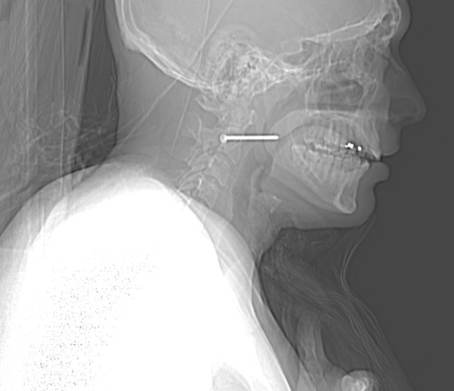
So now what? Well, let’s get some more info! How about a chest xray? Best position? Prone! It’s the easiest, because the patient does not need to be held up next to an xray plate, which would also have to be held manually. The lateral view doesn’t add anything but hassle. Here’s the result:

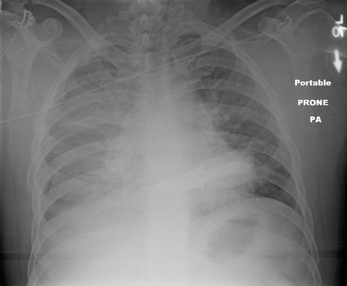
Now what? What do you see, what do you do? Tweet or comment; more to follow tomorrow.

Related post:

What Would You Do? Knife To The Back!

Here’s an interesting case to consider. A young male is assaulted and stabbed to the back. Paramedics bring him to your ED as a trauma team activation, and the full team is assembled prior to his arrival. 

He is brought into the room on the stretcher in the prone position. Here is a representative picture. This is not the actual patient, just a picture I found on another blog site that looks pretty close to the real case.

Let’s walk through the thought processes of managing this over the next few days.

First, what do you need to know right now to navigate your critical decision points? And what are you going to do regarding positioning, evaluation, and imaging?

Tweet or comment with your replies! More on Monday.

Nail In The Neck: Part 2

This case involves an accidental nail gun injury to the neck. The patient is hemodynamically stable, neurologically intact, the airway is patent and not threatened, and there is no apparent hematoma. There is a small puncture near the sternocleidomastoid muscle on the right, fairly high on the neck. The nail is not palpable on either side. And the patient only complains of a little discomfort when he swallows.

What to do? First, the patient has passed all the initial decision points that would send us straight to the OR (ABC problems in ATLS jargon). But, per physical exam and initial imaging, the nail must obviously come out. We just have to figure out what we need to know before we take it out, and determine the best way to retrieve it.

Given the patient’s stability, additional imaging will be helpful. Views in different planes, and details of what the nail might have passed through will be invaluable. The recommended study is a CT angio of the neck. This will give good information about nearby structures and the vasculature. And software reconstructions will provide good 2D/3D information for removal planning. Here’s a lateral view.

image

The nail is located in front of the body of C2. It appears to be high enough to be near the pharynx, but well above trachea and esophagus. You can also see that the nail entered a little posteriorly, and travels right to left and forward. 

Here’s a representative CT slice.

image

The nail enters behind the carotids (just above the bifurcation) and IJ on the right, and ends anterior to them on the left. It passes very close to the posterior pharynx. So neurovascular structures are intact, and the aerodigestive tract is a maybe (back of the pharynx).

Obviously, this thing has to come out. The question is, how to do it? For you surgeons out there, tell me your choice of approach, incision, and instrumentation. Tweet or leave comments! Answers on Monday.

Related posts:

Interesting Case: Part Deux

This is a continuation of yesterday’s interesting case involving an unusual DPL result. As you recall, the tap was negative, but the lavage effluent slowly began to show some particulate material.

By definition, this is a positive result, which then requires a trip to the operating room. The catheter was capped and left in place. The patient was then taken to surgery, prepped and draped. Here’s what was found:

What’s your diagnosis now? And what needs to be done about it?

Final answers tomorrow!

Interesting Case

Here’s an interesting trauma case, which comes from days of DPL. Although we don’t use this valuable technique very often, this one teaches an interesting lesson.

A middle aged female was involved in a high speed car crash. She was brought to the resuscitation bay as a trauma activation because the medics reported she had bilateral femur fractures, and her systolic pressures were in the 90’s. 

As you proceed through the ATLS protocol, you call for blood to supplement your resuscitation fluids, and you also find that her abdomen is tender, with some right upper quadrant guarding. The femurs are placed in traction splints. FAST is generally negative, but the right upper quadrant is equivocal.

At this point, her pressure drops again. You re-evaluate your ABCs and find nothing new. The femurs appear to be nicely reduced, and the thighs are not larger than they were when she arrived. Your surgeon is concerned that the abdomen may be the source despite the (mostly) negative FAST. Due to BP concerns, she proceeds to do a DPL.

The procedure proceeds smoothly while resuscitation with blood products takes place. There is no gross blood on the tap. A liter of saline is infused and is now freely emptying into a bag. For the first 400cc, the effluent is crystal clear. But now you start to see something.

Hmm, is it or isn’t it? Let’s take a closer look.

Yeah, that’s weird. Just of hint of some kind of tiny darkish particles settling to the bottom of the tubing. Hmmm!

So what’s happening here? And what should you do? More information tomorrow. Please comment or tweet your guesses!