Tag Archives: Heart

2 Ways To Perform A Pericardial Window For Trauma: Part 2 With Video!

In my last post, I reviewed the classic, “old-timey” subxyphoid approach to the pericardial window procedure for trauma. Today, I’ll describe the operative approach if you are already in the abdomen managing injuries there.

The same considerations apply to these patients in deciding to perform the window. Either there is a suspicion of actual pericardial tamponade based on physiology or diagnostic imaging, or an injury has been noted in proximity to the heart that raises that suspicion.

If you are already exploring the abdomen, the procedure is much simpler. The instruments required are already in your laparotomy setup:

  • Two toothed forceps
  • Tissue (Metzenbaum) scissors

First, and most importantly, the upper abdomen must be evacuated of all blood. This is critically important since a positive window is solely determined by the presence of blood in the pericardial fluid. If it is contaminated with blood as it flows into the peritoneal cavity, a false positive may result leading to an unneeded thoracotomy or sternotomy.

The midline incision must extend to the xiphoid process in order to get adequate exposure of the diaphragm. The left lobe of the liver is retracted downwards by your assistant, and the two of you can then grasp an area of the pericardial portion of the diaphragm with the toothed forceps. As it is tented away from the heart, the scissors are used to dissect through both the diaphragm and pericardium. Although some use cautery for this, I’m a weenie using electricity near the heart.

The diaphragm is thick, so expect to cut through several mm of tissue before you see pericardial fluid. Watch the color of the fluid carefully. If it is the least bit blood tinged, the result is positive. And be sure to watch for 15-30 seconds. Sometimes the initial fluid is amber, but it becomes bloody as more is drained.

Bloody fluid equals positive result. This means that a thoracic procedure is indicated to evaluate the heart and repair the injury. The choice of sternotomy vs thoracotomy is determined by mechanism, foreign body trajectory, and suspected area of injury on the heart.

If the result is negative, you may close the hole with your suture of choice. If the abdomen is contaminated from a bowel injury, I recommend you use the traditional subxiphoid approach separate from the laparotomy incision to avoid contaminating the pericardial sac.

Here’s a YouTube video of a transdiaphragmatic window created laparoscopically. Since abdominal explorations for major trauma seldom lend themselves to laparoscopy, don’t get any ideas from watching this!

YouTube player

2 Ways To Perform A Pericardial Window For Trauma: Part 1 with Video!

In this two-part post, I’ll describe two ways to perform a pericardial window for trauma. The pericardial window should be considered in any trauma patient who has one of the following:

  • A suspected diagnosis of pericardial tamponade. These patients do not yet have classic signs and symptoms. If they did, a thoracotomy or sternotomy is in order, not a window.  But they do have a mechanism that could produce bleeding into the pericardial sac, and a positive imaging study. Typically, this study is a FAST exam of the heart. Occasionally, pericardial fluid may be seen on chest CT. This is uncommon but significant when detected.
  • An injury in proximity to the heart that is of concern for cardiac injury with a negative or indeterminant FAST. These are typically penetrating injuries so close to the heart that it’s hard to believe it wasn’t injured. If the FAST is not helpful, a window will make the definitive diagnosis.

Pericardial window is a very invasive procedure. For trauma, it is usually performed in the operating room and requires general anesthesia. It could be performed in the ED if the patient is already intubated and sedated.

There are two ways to perform this procedure. Today, I’ll discuss the old-timey subxiphoid approach.  The equipment required is minimal:

  • Scalpel
  • Tissue (Metzenbaum) scissors
  • Once or two toothed forceps
  • Your finger
  • Good lighting

A 4-8 cm incision is made extending from the top of the xiphoid, extending about 4 cm down onto the abdominal midline. Enter the retrosternal space with your finger, and head to the heart. Usually, some fatty tissue must be bluntly dissected out of the way. Note: the heart is frequently further away than you think!

Sweep the fat out of the way, exposing the pericardium. Grasp the pericardium with the toothed forceps and tent it away from the heart. Use the Metzenbaum scissors to incise the pericardium immediately adjacent to the forceps. If this is difficult, then have an assistant grasp the pericardium with another pair so a short line of pericardium is elevated. (Note: sometimes having a second set of forceps in the incision makes it too difficult to see, which is why I prefer the single forceps technique).

Make sure that the wound is bloodless when you incise the pericardium! There is always at least a small amount of pericardial fluid that will squirt out, and you are looking at its color. If it is anything but amber, you have a positive result. If you have a bloody field that contaminates the fluid, a false positive diagnosis could occur leading to an unnecessary thoracotomy.

If the window is positive, cover the wound and head immediately to the OR if your’re not already there. Your patient has a cardiac injury until proven otherwise. If negative, then close the skin wound with your sutures / staples of choice. Do not attempt to close the tiny pericardial hole!

Here’s a video that shows the basic technique. The procedure depicted is being performed for non-trauma, so the operator takes his time. He has the luxury of dissecting and exposing the field well. But in trauma, we don’t usually have time to resect the xiphoid or take 10 minutes to dissect out the field.

YouTube player

In my next post, I’ll discuss the technique that is used if you already find yourself in the abdomen when a cardiac injury is suspected.

Off-Label Use of the Foley (Urinary) Catheter

Foley catheters are a mainstay of medical care in patients who need control or measurement of urine output. Leave it to trauma surgeons to find warped, new ways to use them!

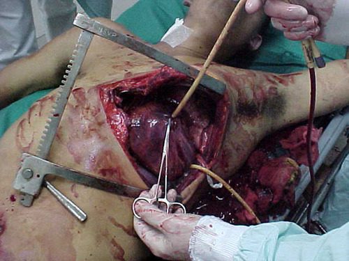
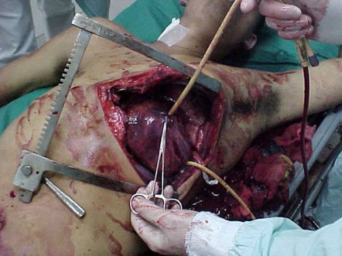
Use of these catheters to tamponade penetrating cardiac injuries has been recognized for decades (see picture, 2 holes = 2 catheters!). Less well appreciated is their use to stop bleeding from other penetrating wounds.

Foley catheters can be inserted into just about any small penetrating wound with bleeding that does not respond to direct pressure. (Remember, direct pressure is applied by one or two fingers only, with no flat dressings underneath to diffuse the pressure). Arterial bleeding, venous bleeding or both can be controlled with this technique.

In general, the largest catheter with the largest possible balloon should be selected. It is then inserted directly into the wound until the entire balloon is inside the body. Inflate the balloon using saline until firm resistance is encounted, and the bleeding hopefully stops. Important: be sure to clamp the end of the catheter so the bleeding doesn’t find the easy way out!

Use of catheter tamponade buys some time, but these patients need to be in the OR. In general, once other life threatening issues are dealt with in the resuscitation room, the patient should be moved directly to the operating room. In rare cases, an angiogram may be needed to help determine the type of repair. However, in the vast majority of cases, the surgeon will know exactly where the injury is and further study is not needed. The catheter is then prepped along with most of the patient so that the operative repair can be completed.

Tomorrow: an off-label use for this catheter in abdominal trauma!

Off-Label Use of the Foley Catheter

Foley catheters are a mainstay of medical care in patients who need control or measurement of urine output. Leave it to trauma surgeons to find warped, new ways to use them!

Use of these catheters to tamponade penetrating cardiac injuries has been recognized for decades (see picture, 2 holes!). Less well appreciated is their use to stop bleeding from other penetrating wounds.

foleyinheart

Foley catheters can be inserted into just about any small penetrating wound with bleeding that does not respond to direct pressure. (Remember, direct pressure is applied by one or two fingers only, with no flat dressings underneath to diffuse the pressure). Arterial bleeding, venous bleeding or both can be controlled with this technique.

In general, the largest catheter with the largest possible balloon should be selected. It is then inserted directly into the wound until the entire balloon is inside the body. Inflate the balloon using saline until firm resistance is encounted, and the bleeding hopefully stops. Important: be sure to clamp the end of the catheter so the bleeding doesn’t find the easy way out!

Use of catheter tamponade buys some time, but these patients need to be in the OR. In general, once other life threatening issues are dealt with in the resuscitation room, the patient should be moved directly to the operating room. In rare cases, an angiogram may be needed to help determine the type of repair. However, in the vast majority of cases, the surgeon will know exactly where the injury is and further study is not needed. The catheter is then prepped along with most of the patient so that the operative repair can be completed.

Related posts: