Tag Archives: gunshot

Can Lead Poisoning Occur After A Gunshot?

This is a fairly common question from victims of gunshots and their families. As you know, bullets are routinely left in place unless they are superficial. It may cause more damage to try to extract one, especially if it has come to rest in a deep location. But is there danger in leaving the bullet alone?

One of the classic papers on this topic was published in 1982 by Erwin Thal at Parkland Hospital in Dallas. The paper recounted a series of 16 patients who had developed signs and symptoms of lead poisoning (plumbism) after a gunshot or shotgun injury. The common thread in these cases was that the injury involved a joint or bursa near a joint. In some cases the missile passed through the joint/bursa but came to rest nearby, and a synovial pseudocyst formed which included the piece of lead. The joint fluid bathing the projectile caused lead to leach into the circulation.

The patients in the Parkland paper developed symptoms anywhere from 3 days to 40 years after injury. As is the case with plumbism, symptoms were variable and nonspecific. Patients presented with abdominal pain, anemia, cognitive problems, renal dysfunction and seizures to name a few.
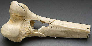
Bottom line: Any patient with a bullet or lead shot that is located in or near a joint or bursa should have the missile(s) promptly and surgically removed. Any lead that has come to rest within the GI tract (particularly the stomach) must be removed as well. If a patient presents with odd symptoms and has a history of a retained bullet, obtain a toxicology consult and begin a workup for lead poisoning. If levels are elevated, the missile must be extracted. Chelation therapy should be started preop because manipulation of the site may further increase lead levels. The missile and any stained tissues or pseudocyst must be removed in their entirety.

Reference: Lead poisoning from retained bullets. Ann Surg 195(3):305-313, 1982.

The Cardiac Box: Meaningful For Gunshot Wounds?

A common dogma in trauma training is: “Watch out for the box!” This area on the anterior chest is purported to indicate high risk of cardiac injury in patients with penetrating trauma.

Where is it, exactly? Technically, it’s the zone extending from nipple to nipple, and from sternal notch to xiphoid.

The cardiac box

But is the dogma true? A number of (old) papers mapped out the location and incidence of cardiac injury in stabs to the chest and upper abdomen. And there is a pretty good correlation. For stab wounds. But what about gunshots?

A team at Emory University ran a retrospective review of their trauma registry data over a three year period.

Here are the factoids:

  • They saw nearly 90 patients per year with penetrating chest wounds. Of these, 80% were gunshots (!) Many had more than one penetration.
  •  Of the 233 gunshots inside “the box”, 34% injured the heart
  • The remaining 44 gunshots outside “the box” hit the heart 32% of the time
  • The authors suggest shifting the definition of “the box” toward the left, so that it extends from anterior midline, wraps around the left chest, and ends in the posterior midline (see below)

new-cardiac-box

Bottom line: Here’s the problem. Knives are attached to a handle which tends to stay outside your patient. Thus, it can only go so deep. But a bullet will keep going until something stops it, or it runs out of gas. So it makes sense that the traditional boundaries of “the box” don’t apply. But extending it to include the left lateral chest and exclude everything on the right side? It may make statistical sense in this study, but common sense dictates that the trauma professional needs to think about the heart any time a gunshot goes anywhere near the chest or upper abdomen. Do not limit yourself to any “box!”

Reference: Redefining the cardiac box: evaluation of the relationship between thoracic gunshot wounds and cardiac injury. AAST 2016 Paper #12.

Can Lead Poisoning Occur After A Gunshot?

This is a fairly common question from victims of gunshots and their families. As you know, bullets are routinely left in place unless they are superficial. It may cause more damage to try to extract one, especially if it has come to rest in a deep location. But is there danger in leaving the bullet alone?

One of the classic papers on this topic was published in 1982 by Erwin Thal at Parkland Hospital in Dallas. The paper recounted a series of 16 patients who had developed signs and symptoms of lead poisoning (plumbism) after a gunshot or shotgun injury. The common thread in these cases was that the injury involved a joint or bursa near a joint. In some cases the missile passed through the joint/bursa but came to rest nearby, and a synovial pseudocyst formed which included the piece of lead. The joint fluid bathing the projectile caused lead to leach into the circulation.

The patients in the Parkland paper developed symptoms anywhere from 3 days to 40 years after injury. As is the case with plumbism, symptoms were variable and nonspecific. Patients presented with abdominal pain, anemia, cognitive problems, renal dysfunction and seizures to name a few.

Bottom line: Any patient with a bullet or lead shot that is located in or near a joint or bursa should have the missile(s) promptly and surgically removed. Any lead that has come to rest within the GI tract (particularly the stomach) must be removed as well. If a patient presents with odd symptoms and has a history of a retained bullet, obtain a toxicology consult and begin a workup for lead poisoning. If levels are elevated, the missile must be extracted. Chelation therapy should be started preop because manipulation of the site may further increase lead levels. The missile and any stained tissues or pseudocyst must be removed in their entirety.

Reference: Lead poisoning from retained bullets. Ann Surg 195(3):305-313, 1982.

Gunshots And CT Scan Of The Abdomen

Abdominal gunshots and CT scanning are usually thought to be mutually exclusive. The usual algorithm generally means a prompt trip to the operating room. But as with many things in the management of trauma, there are always exceptions. The key is to understand when exactly one of those exceptions is warranted.

Exception 1: Did it really enter the abdomen? Gunshots have enough energy that they usually do get inside. However, freaky combinations of trajectory and body habitus do occur. There are three tests that must be passed in order to entertain the possibility that the bullet may not have made it inside your patient: physiology, anatomy, and physical exam. For physiology, the patient must be completely hemodynamically stable. Anatomically, the trajectory must make sense. If the known wounds and angles allow a tangential course make sense, then fine. But if there is a hole in the epigastrium and another next to the spine, you have to assume the bullet went straight through. Finally, the physical exam must be normal. No peritonitis. No generalized guarding. Focal tenderness only in the immediate area of any wounds. If all three of these criteria are passed, then a CT can be obtained to demonstrate the trajectory.

Exception 2: Did it enter an unimportant area of the abdomen? Well, there’s really only one of these, and that’s the area involving the right lobe of the liver and extending posteriorly and lateral to it. If the bullet hole(s) involve only this area, and the three tests above are passed, CT may confirm an injury that can be observed. However, there should only be a minimal amount of free fluid, and no soft tissue changes of any kind adjacent to bowel.

Exception 3: A prompt trauma lap was performed, but you think you need more information afterwards. This is rare. The usual belief is that the eyes of the surgeon provide the gold standard evaluation during a trauma lap. For most low velocity injuries with an easily understood trajectory, this is probably true. However, high velocity injuries, those involving multiple projectiles, or complicated trajectories (side to side) can be challenging for even the most experienced surgeon. Some areas (think retroperitoneum or deep in the pelvis) are tough to visualize completely, especially when there’s blood everywhere. These are also the cases most likely to require damage control surgery, so once the patient has been temporarily closed, warmed and resuscitated, a quick trip to CT may be helful in revealing unexpected shrapnel, unsuspected injuries, or other issues that may change your management. Even a completely unsurprising scan can provide a higher sense of security.

Bottom line: CT of the abdomen and gunshots to that area may actually coexist in some special cases. Make sure the physiology, anatomy and physical exam criteria are passed first. I also make a point of announcing to all trainees that taking these patients to CT is not the norm, and carefully explain the rationale. Finally, apply the concept of the null hypothesis to this situation. Your null hypothesis should state that your patient does not need a CT after gunshot to the abdomen, and you have to work to prove otherwise!

Penetrating Injuries to the Extremities

Simple penetrating injuries to the arms and legs are often over-treated with invasive testing and admission for observation. Frequently, these injuries can be rapidly evaluated and disposed of using physical examination skills alone.

Stabs and low velocity gunshots (no rifles or shotguns, please) should be thoroughly examined. This includes an examination of the entire, unclothed body. If this is not carried out, there is a risk that additional penetrating injuries may be missed.

For gunshots, look at the wounds and the estimated trajectory to try to demonstrate that the object stayed clear of neurovascular structures. This exam is imprecise, and must be accompanied by a full neurovascular exam and evaluation of the bones and joints. If there is any doubt regarding bony involvement, plain radiographs with entry markers should be performed. Any abnormal findings will require more in-depth evaluation and inpatient admission.

If the exam is negative but the trajectory is “in proximity” to a major vessel, an arterial pressure index (API) should be measured. This test involves the calculation of the ratio of the systolic pressure in the injured extremity to the contralateral uninjured extremity. It should not be confused with the ankle brachial index (ABI) which compares the systolic pressure in the ipsilateral uninjured arm  or leg.

The magic ratio is 0.9. If the API is less than this, there is some likelihood that a vascular injury is present. If the API is higher, there is virtually no chance of injury.

The final test that must be performed before discharge is a function test. If the injured extremity is too painful to use or walk on, the patient may need to be admitted for pain management and therapy. Patients managed in this way can avoid arteriography, CT angiography or admission and save thousands of dollars in hospital charges.

Reference: Journal Am Coll Surgeons 2009;209:740-5.