Tag Archives: fasciotomy

Once Again: Leg Fasciotomy With Less Than Two Incisions

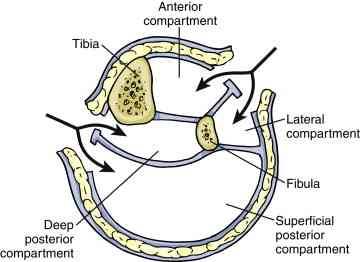
It seems like this topic keeps on coming up! This is the second article I’ve seen this year that describes a variation on the single-incision leg fasciotomy. In the classic two-incision approach, the lateral incision gives access to the anterior and lateral compartments, and the medial incision to the posterior and deep posterior. See below.

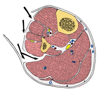
The more “standard” single-incision approaches either go through (i.e. removes part of) or around, the fibula. In the diagram below, the arrows point to the access points into the anterior, lateral, posterior and deep posterior from top to bottom.

In the “new” variation described, the authors slide along the lateral edge of the tibia to get to the deep posterior compartment.

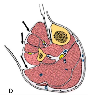
This approach requires stripping the tibialis

anterior muscle away from the tibia, which some orthopods may argue interferes with healing. And, as with the other single-incision technique, the procedure may take additional time.

Bottom line: I’m still not a big fan of single-incision fasciotomy. My main reason is that most surgeons are not as familiar with the technique. And patients who have a potentially limb threatening process are not the best to learn on. I have seen too many incomplete fasciotomies with persistent compartment syndrome in my career. 

So unless you are being mentored by someone who is well versed in the technique, use the two incision technique and use a cadaver to practice your single incision operation.

Reference:  A Single-Incision Fasciotomy for Compartment Syndrome
of the Lower Leg. J Ortho Surg 30(7):e252-e255, 2016.

Single Incision Fasciotomy?

The concept of performing 4-compartment leg fasciotomy using only one incision is not a new one. Techniques using a lateral approach, either with or without fibulectomy, have been described as early as 1967.

A new paper describes a single-incision technique for compartment syndrome using a medial approach. The authors believe that going through the anterior compartment to release the deep posterior is quicker, uses a smaller flap, and avoids injury to the peroneal nerve. They reviewed their own experience over a 5 year period.

Here are the factoids:

  • 180 fasciotomies were performed for compartment syndrome, of which 30 were single-incision
  • 27 had associated fractures, 2 were due to soft tissue injury and 1 was spontaneous
  • There was a single wound infection, nerve injury, and patient with persistent pain. There were several tethered tendons and scars.
  • The number and types of complications were similar to traditional fasciotomy

Traditional 4 compartment fasciotomy with 2 incisions. Source: gog.net.nz

Bottom line: Sounds great, right? Yes, it’s a small study, and statistically there is not enough power to show that it’s “better.” So if it’s not worse, and there is just one smaller incision, what’s wrong with it?

For me, the problem is that there is too much opportunity to perform an incomplete fasciotomy. The learning curve for single incision fasciotomy, either this one or the more traditional lateral approach, is steep. Seriously impaired patients who need fasciotomy are frequently not going to be awake any time soon, leaving the surgeon with no neurologic or pain exam.

My recommendation: read papers like this and smile. Then do the classic 2 incision operation, making sure that all compartments are completely released.

Related post:

Reference: A single incision fasciotomy for compartment syndrome of the lower leg. J Ortho Trauma, Publish ahead of print, January 21, 2016.

New Technique for Fasciotomy Closure

Fasciotomies are much more easily opened than closed! Once the edematous muscle is released, it’s not easy to get the skin to close over it again. On occasion, an immediate closure can be carried out. But in most cases, the process is performed with one or more additional operations.

Continuous tension across the skin edges is important. This keeps the wound from getting wider while the edema decreases. A number of creative techniques have been employed to keep the wound from widening, including using sutures, vessel loops, and fancy (expensive) plastic fasteners. And although the KCI VAC dressing reduces edema, it does not do much to pull the wound edges together.

Surgeons in the Netherlands came up with a novel technique using a cheap device that can be found in any hardware store and gas sterilized. The Ty-Rap closure device is commonly used to secure chest tubes to their connectors. Bigger versions are used by police in lieu of handcuffs.

The tail of one Ty-Rap is cut off and the hub is placed on the tail end of another. This assembly is placed across the wound, and one staple is placed over it on each side of the wound. This process is repeated for the entire length of the wound (picture). The Ty-Raps are tightened, and then slowly retightened daily until the wound comes together. An additional week to 10 days is allowed for wound healing before removal of the Ty-Raps.

The authors used this technique on 23 extremity fasciotomy wounds. The wounds were closed after an average of 6 days, and the TyRaps were removed after 16 days. There was no skin necrosis, but there were two instances of cellulitis. The cost of the materials (TyRaps and a surgical stapler) was $23, excluding assembly and sterilization.

Bottom line: This is an interesting technique with good closure results. The surgeon does have to plan ahead and get hospital clearance to use these devices, though.

Reference: Ty-Raps in trauma:  a novel closing technique of extremity fasciotomy wounds. J Trauma 69:972-975, 2010.

TyRap closure