Tag Archives: DPL

Interesting Case: Part Deux

This is a continuation of yesterday’s interesting case involving an unusual DPL result. As you recall, the tap was negative, but the lavage effluent slowly began to show some particulate material.

By definition, this is a positive result, which then requires a trip to the operating room. The catheter was capped and left in place. The patient was then taken to surgery, prepped and draped. Here’s what was found:

What’s your diagnosis now? And what needs to be done about it?

Final answers tomorrow!

Interesting Case

Here’s an interesting trauma case, which comes from days of DPL. Although we don’t use this valuable technique very often, this one teaches an interesting lesson.

A middle aged female was involved in a high speed car crash. She was brought to the resuscitation bay as a trauma activation because the medics reported she had bilateral femur fractures, and her systolic pressures were in the 90’s. 

As you proceed through the ATLS protocol, you call for blood to supplement your resuscitation fluids, and you also find that her abdomen is tender, with some right upper quadrant guarding. The femurs are placed in traction splints. FAST is generally negative, but the right upper quadrant is equivocal.

At this point, her pressure drops again. You re-evaluate your ABCs and find nothing new. The femurs appear to be nicely reduced, and the thighs are not larger than they were when she arrived. Your surgeon is concerned that the abdomen may be the source despite the (mostly) negative FAST. Due to BP concerns, she proceeds to do a DPL.

The procedure proceeds smoothly while resuscitation with blood products takes place. There is no gross blood on the tap. A liter of saline is infused and is now freely emptying into a bag. For the first 400cc, the effluent is crystal clear. But now you start to see something.

Hmm, is it or isn’t it? Let’s take a closer look.

Yeah, that’s weird. Just of hint of some kind of tiny darkish particles settling to the bottom of the tubing. Hmmm!

So what’s happening here? And what should you do? More information tomorrow. Please comment or tweet your guesses!

DPL: A Dying Art?

Diagnostic peritoneal lavage (DPL) was invented by David Root at this hospital (Ancker Hospital, which then became St. Paul Ramsey, now Regions Hospital) in the 1960’s. It enjoyed its heyday during the 70’s and 80’s, when it was done hundreds of times per year at most major trauma hospitals. Now, we do it about 5 times per year. What happened?

As you know, DPL is a qualitative test. It gives a yes/no answer to the question “does this patient need an operation?” based on red and white blood cell counts. During the mid-1980s, CT scanning was introduced, which provides much more quantitative information about injuries in the abdominal cavity. The improved ability to diagnose abdominal injury, especially solid organ injury, has led to the demise of DPL.

Most solid organ injury results in some free blood in the peritoneal cavity. It doesn’t take much blood (10 cc of whole blood mixed with 1 liter of infused crystalloid) to exceed the threshold of 100,000 RBC per ml of aspirate that will send the surgeon off to the OR. Therefore, pretty much any liver or spleen laceration would have to be taken to the OR based on a DPL.

But we know that very few liver/spleen injuries actually need an operation. So DPL cannot be used, or the negative laparotomy rate for blunt trauma would escalate. The other downside to DPL is that it’s not possible to get all of the infused crystalloid back out of the abdomen. This leads to a confusing amount of free fluid seen on any CT scan done after a DPL.

So DPL is now down but not out. Some practical pointers:

  • DPL should be used primarily as a backup to an equivocal or unbelievable FAST exam in an unstable patient. An example would be a patient who is hypotensive, has a negative FAST and no other obvious bleeding sources.
  • Remember to insert a gastric tube and urinary catheter so the stomach and bladder are decompressed before the procedure. The easiest way to remember this is to tape these catheters to the DPL procedure tray.
  • A DPL is actually 2 procedures: peritoneal tap and lavage. Once the catheter is in, it should be aspirated. If 10cc of gross blood is returned, the test is positive and the patient needs to go immediately to OR.
  • For blunt trauma, the threshold for RBC per µl is 100,000. The threshold for WBC is 500 per µl. If particulate material or weird colors are seen (stool or bile), the test is also considered positive. Send the sample for cell counts only. Don’t send for any other assays (e.g. amylase). 
  • For penetrating trauma, the thresholds have never been well defined. A number around 25,000 RBC per µl probably provides the best balance between sensitivity and negative laparotomy rate.

Reference: Diagnostic peritoneal lavage. HD Root, CW Hauser, CR McKinley, JW LaFave, RP Mendiola Jr. Surgery 57(5):633-637, 1965.

Pop Quiz! DPL – The Answer!

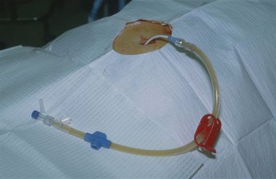
You’re doing one of those (very rare) DPLs and get a surprise result. Not blood, not obvious intestinal content, but just a small amount of mysterious sediment. What to do?

Well, this is obviously not normal. Therefore, this has to be considered a positive diagnostic peritoneal lavage. Since DPL is a qualitative test (meaning that the answer is only yes or no), the patient must go to the OR.

Here are the answers to the questions posed earlier today:

  • The DPL catheter has a relatively small diameter, so leave it in place! It may be very difficult to find where it went otherwise
  • Midline laparotomy incision is most appropriate. Remember, this is a trauma case? However, you can start infra-umbilical with a limited incision.

Here’s what I found in this case:

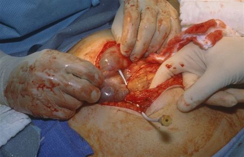
The catheter went straight into the cecum! So we actually did a diagnostic colonic lavage! The sediment was a very small amount of stool. And as stated above, had the catheter not been left in place, it would have been very tough to find the puncture site. 

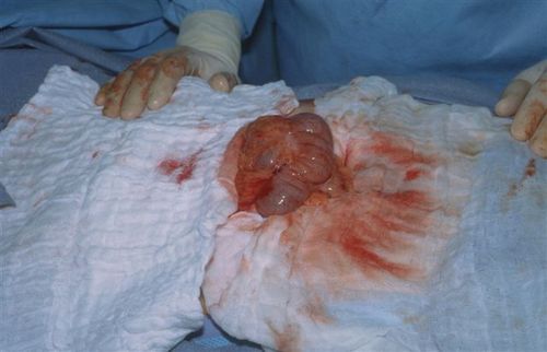
Next, I clamped the catheter to keep it in place, cut it on the hub side, and removed most of it.

Finally, I placed a purse-string stitch around the entry site in the bowel, removed the catheter and tied the suture.

But wait, we’re not done yet! The patient did have abdominal pain and a seat belt sign, so we did a trauma exploration through the midline incision. A Grade II liver injury was present which needed no further management. The patient did well  and was discharged on the fourth day.

Bottom line: Procedures can and do go awry. Reason your way through it the best you can, then use focused diagnostics, if needed, to come up with a plan. For misplaced needles and catheters, most organs can tolerate a puncture by almost anything (except the eye, maybe). Treat appropriately and monitor carefully afterwards.

Source: Personal archive. Not treated at Regions Hospital

Pop Quiz! DPL Hint

So the catheter is in, the aspirate was negative (nothing came out), and a liter of crystalloid infused easily. But toward the end of draining the fluid back out, some faint sediment became visible in the tubing.

A lot of you guessed bladder, but most people don’t have sediment there. Plus, if I dumped a liter of fluid into your bladder, you’d really get the urge to go. This awake patient noted no new symptoms. 

I had a bad feeling about this, so I elected to take her to the OR to see what the story really was. Here are some questions for any budding surgeons out there:

  • Leave the catheter in place or pull it out before OR?
  • What incision to make?
  • How big?
  • And what the heck is it, really?

Answers later today! See if you can get it before I give you the punch line!