Tag Archives: damage control

A Sample Final Damage Control X-ray

Yesterday, I wrote about ways to reduce and hopefully eliminate retained foreign bodies (instruments, sponges) during damage control surgery. Today, I’ll provide a sample x-ray and some tips on how to use this tool most effectively.

Here is an abdominal x-ray obtained just prior to closure of a patient who underwent damage control laparotomy. The OR record and surgeon from the initial operation documented that four sponges had been left in place for hemostasis.
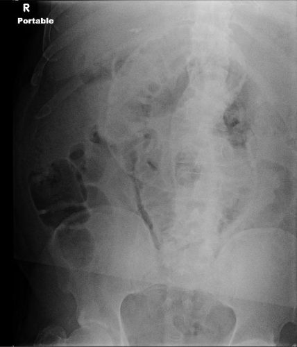
dc-closure

Nothing retained, right?

Wrong! This image is not complete. This patient is larger than the x-ray plate used. The area under the diaphragms, the pelvis, and the entire left side of the peritoneal cavity have not been visualized.

Tips for imaging for damage control closure:

  • Always make sure the patient is on an x-ray OR table. It is so annoying (and potentially a sterility problem) to have to slide the plate under the patient!
  • Help the radiology tech to locate the desired imaging field using folds in the towels covering the body region. For example place the confluence of folds in the center of the towel in the exact place you want the center of the x-ray to be.
  • Remove all radiopaque objects from the x-ray field to reduce confusion when interpreting the image
  • Make sure the entire body cavity has been imaged! This may mean bracketing the area with several shots.
  • Read the image yourself! But if in doubt, or in patients with drains or other odd objects, call the radiologist to help you out.

Related posts:

The Final X-Ray In Damage Control Surgery

Damage control surgery for trauma is over 20 years old, yet we continue to find ways to refine it and make it better. Many lives have been saved over the years, but we’ve also discovered new questions. How soon should the patient go back for definitive closure? What is the optimal closure technique? What if it still won’t close?

One other troublesome issue surfaced as well. We discovered that it is entirely possible to leave things behind. Retained foreign bodies are the bane of any surgeon, and many, many systems are in place to avoid them. However, many of these processes are not possible in emergent trauma surgery. Preop instrument counts cannot be done. Handfuls of uncounted sponges may be packed into the wound.

I was only able to find one paper describing how often things are left behind in damage control surgery (see reference below), and it was uncommon in this single center study (3 cases out of about 2500 patients). However, it can be catastrophic, causing sepsis, physical damage to adjacent organs, and the risk of performing an additional operation in a sick trauma patient.

So what can we do to reduce the risk, hopefully to zero? Here are my  recommendations:

  • For busy centers that do frequent laparotomy or thoracotomy for trauma and have packs open and ready, pre-count all instruments and document it
  • Pre-count a set number of laparotomy pads into the packs
  • Use only items that are radiopaque or have a marker embedded in them. This includes surgical towels, too!
  • Implement a damage control closure x-ray policy. When the patient returns to OR and the surgeons are ready to begin the final closure, obtain an x-ray of the entire area that was operated upon. This must be performed and read before the closure is complete so that any identified retained objects can be removed.

Tomorrow, a sample damage control closure x-ray.

Related post:

Reference: Retained foreign bodies after emergent trauma surgery: incidence after 2526 cavitary explorations. Am Surg 73(10):1031-1034, 2007.

Damage Control Dressing: The ABThera (Video)

In the late 1980’s, when we started the work that would be published in the first damage control paper from Penn, we used the vacuum pack dressing. This was first described in a paper from the University of Tennessee at Chattanooga in 1995. Prior to that, the so-called Bogota bag was the usual technique. This consisted of slicing opening up a sterile IV bag (either the standard 1 liter or the urology 3 liter bag for big jobs) and sewing it into the wound. This worked, but it freaked out the nurses, who could see the intestines through the print on the clear plastic bag.

The vacuum pack was patient friendly, with a layer of plastic on the bottom, some absorbent towels in the middle with a drain in place to remove fluid and apply suction, and an adherent plastic layer on top to keep the bed clean. As you can imagine, this was a little complicated to apply correctly. One misstep and things stuck to the bowel or leaked out onto the bed.

In the past few years, a commercial product was developed that incorporated all these principles and was easy to apply. This is the KCI ABThera (note: I have no financial interest in KCI or this product; I just wish I had invented it). The only downside is that there is a small learning curve when first using this product.

YouTube player

The video above shows a demonstration of the application on an abdominal mannikin. It is not as slick as the company videos, but I think it’s more practical, with some good tips.

References:

  • Damage control: an approach for improved survival in exsanguinating penetrating abdominal injury. J Trauma 35(3):375-382, 1993.
  • Temporary closure of open abdominal wounds. Am Surg 61(1):30-35, 1995.

Prone Positioning After Laparotomy

Patients with serious abdominal injury may require a laparotomy, and a subset of these may need a temporary closure for damage control surgery. Concomitant spine injury may have your spine surgeons asking “is it safe to prone the patient who is postop with a midline incision or an open abdomen.” What to tell them?

There’s not much guidance out there in the literature. One paper from 2000 looked at four patients who were proned for severe ARDS and found that one suffered a wound dehiscence. However, this patient had severe generalized edema and was on several pressor agents. 

The use of temporary abdominal closure techniques has revolutionized the early management of severely injured trauma patients and has greatly decreased the incidence of complications from abdominal compartment syndrome. Several authors have now demonstrated that putting those patients in the prone position is well tolerated. 

As far as patients who have a closed laparotomy, proning appears to be well tolerated as well. One caveat: consider carefully if the patient is having wound complications or if they are morbidly obese.

The bottom line: Consider the risks and benefits carefully in any post-laparotomy patient you are considering prone positioning for. Other than in morbidly obese, it is generally considered safe, even in patients with damage control dressings in place. However, make sure the trauma surgeon re-evaluates the wound again as soon as the patient is returned to the supine position.

References: 

1. The “open abdomen” is not a contra-indication to prone positioning for severe ARDS (abstract). Schwab, et al. Chest. 1996;110:142S.

2. Complications of Prone Ventilation in Patients with Multisystem Trauma with Fulminant Acute Respiratory Distress Syndrome. Offner et al. Journal of Trauma-Injury Infection & Critical Care. 48(2):224-228, February 2000.

3. The Management of the Open Abdomen in Trauma and Emergency General Surgery: Part 1-Damage Control. Diaz et al. Journal of Trauma-Injury Infection & Critical Care. 68(6):1425-1438, June 2010.