Tag Archives: CT

What The Heck? CT Imaging Problem: The Answer

I received some good guesses about this image yesterday, but no one got the right answer.

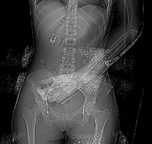
The patient had sustained blunt trauma and was undergoing CT imaging. The scout for the abdominal CT showed some kind of weird debris that interfered with the image, but when we uncovered and looked at the patient, nothing was visible:

What the heck? If you look carefully at the left side of the image, you can see that the “debris field” is on the surface of the patient. We can’t see in 3-D on images, but the difference in appearance on the left and right sides looks like it this stuff is wrapping around the patient.

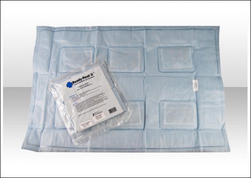
She was brought in by EMS with a warming blanket in place. On closer inspection, this was a thin, disposable blanket that heats up when removed from an airtight plastic pouch. These blankets contain thin pockets of a mineral mixture that looks like gravel. When exposed to air it heats up.

But on CT it looks like bone density material! When we looked at the patient, we were just lifting off the blanket that contained the offending material. Hence, we couldn’t find it.

Here’s a picture of one of these products. Note the six mineral pouches embedded in it., Don’t let this happen to you!


What The Heck? CT Imaging Problem

Here’s one for you. A patient is brought to you after a motor vehicle crash. You’ve completed your evaluation in the trauma resuscitation room, and you move off to CT for some imaging.

As the techs are preparing to do the abdominal CT, they perform the scout image to set up the study. This is what you see:

The arm was left down due to a fracture (note the splint along the forearm). But what is all that debris on the image? Other than a few abrasions here and there, nothing is visible on the skin in those areas.

What the heck? What do you think these are? Will they interfere with imaging? And what can you do about it?

Tweet or comment with your answers. I will explain all tomorrow.

IV Contrast and Trauma – Revisited

We use CT scanning in trauma care so much that we tend to take it (and its safety) for granted. I’ve written quite a bit about thoughtful use of radiographic studies to achieve a reasonable patient exposure to xrays. But another thing to think about is the use of IV contrast.

IV contrast is a hyperosmolar solution that contains some substance (usually an iodine compound) that is radiopaque to some degree. It has been shown to have a significant impact on short-term kidney function and in some cases can cause renal failure.

Here are some facts you need to know:

  • Contrast nephrotoxicity is defined as a 25% increase in serum creatinine, usually within the first 3 days after administration
  • There is usually normal urine output and minimal to no proteinuria
  • In most cases, renal function returns to normal after 3-4 days
  • Nephrotoxicity almost never occurs in people with normal baseline kidney function
  • Large or repeated doses given within 72 hours greatly increase risk for toxicity
  • Old age and pre-existing diabetic renal impairment also greatly increase risk

If you must give contrast to a patient who is at risk, make sure they are volume expanded (tough in trauma patients), or consider giving acetylcysteine or using isosmolar contrast (controversial, may still cause toxicity).

Bottom line: If you are considering contrast CT, try to get a history to see if the patient is at risk for nephrotoxicity. Also consider all of the studies that will be needed and try to consolidate your contrast dosing. For example, you can get CT chest/abdomen/pelvis and CT angio of the neck with one contrast bolus. Consider low dose contrast injection if the patient needs formal angiographic studies in the IR suite. And finally, consider what changes will be made if the study is positive. For example, if a CT angio of the neck for blunt carotid/vertebral injury is being considered, the intervention for a positive result is usually just aspirin. Since this is a very benign medication, why not forgo the scan and just start aspirin if there is a significant risk of kidney injury from the contrast. Always think about the global needs of your patient and plan accordingly (and safely).

Reference: Contrast media and the kidney. British J Radiol 76:513-518, 2003.

Best of AAST #5: Pneumothorax – How Big Is Too Big?

Deciding when to place a chest tube can be challenging. Sometimes, it’s obvious: there is a large hemo- or pneumothorax staring you in the face on the chest x-ray. But sometimes, it’s there but “not that big.” The real question is, how big is too big.

That’s a question that’s been very difficult to quantify. The authors of this abstract, from the Medical College of Wisconsin, conducted a six-year retrospective review of every patient with an isolated pneumothorax at their Level I trauma center. Based on their previous research, a 35mm threshold was used to stratify patients into two groups. This measurement was obtained from axial images of a CT scan. Statistical analysis was performed to identify the predictive value in determining whether the patient could be managed without a chest tube.

Here are the factoids:

  • A total of 1767 patients had a pneumothorax during the 6-year period, and about half met inclusion criteria for the study
  • Of the 385 with pneumothorax alone, 92% were managed without a chest tube
  • Of those 353, 95% had a maximum chest wall to lung distance (335)
  • The 35mm measurement was statistically shown to be an independent predictor of successful management without a tube for both blunt and penetrating trauma

Bottom line: Not so fast! Although this looks like a slam dunk abstract, it’s really not. First, many (or most?) pneumothoraces are initially diagnosed using a plain old chest x-ray. A 35mm measurement is meaningless here because there can be significant changes in position of the pneumothorax on the image. Sometimes, the air is located anteriorly with little or no lateral component.  Does this mean we should CT every patient with a known or suspected pneumothorax? I think not.

And the second issue is the subjectivity surrounding the definition of a failure. What criteria were used when the tube was actually placed in this series. If every patient had to become symptomatic first, then I might agree. But I suspect the tubes were placed when followup imaging showed that the air was just “too big.” You can’t statistic away this kind of potential bias from subjectivity.

So what’s the answer? Unfortunately, there still isn’t one. The need for a chest tube must still be based on subjective size on a chest x-ray, physiologic status, and the patient’s ability to tolerate a given amount of lost lung function. It continues to boil down to the  assessments of each trauma professional as to “how big is too big.”

Reference: Observing pneumothoraces: the 35mm rule is safe for both blunt and penetrating chest trauma. Session XVA Paper 28, AAST 2018.
Torso Trauma CT (Nearly) ALWAYS Requires Contrast

Most stable patients with blunt trauma undergo CT scanning these days. Hopefully, it’s done thoughtfully to optimize the risk/benefit ratio using a well-designed imaging protocol. The majority of these torso imaging protocols call for the use of IV contrast. But as I’ve written before, this can pose risks, especially to the elderly and others who have some degree of renal impairment.

Unfortunately, I occasionally encounter scans done at other hospitals that omit the use of contrast. This usually hinders diagnosis significantly. And it’s usually not clear why this happened, so let’s think about it a bit.

The use of contrast in CT is designed to show blood, or things that are filled with lots of blood. Specifically, a great deal of detail about the blood vessels and solid organs is displayed.

Let’s break it down by type of scan:

  • Chest – we are really only interested in the aorta. The only way to reliably demonstrate an aortic injury is by using contrast. And this is one of those injuries that, if you miss it, the patient is very likely to die from it. Therefore, if you are ordering a chest CT properly, you must add contrast.
  • Abdomen/pelvis – generally, we are looking for solid organ injury, potential mesenteric injuries, and extravasation of blood from organs or soft tissue. Once again, the only way to really see any of these is with contrast enhancement.
  • Vascular – CT is replacing conventional angiography for the investigation of vascular injury in many cases. Obviously, this study is worthless without the contrast.

Bottom line: Pretty much any CT of the chest, blood vessels, or abdomen/pelvis must have IV contrast injected for accurate diagnosis. But what if your patient is old, or is known to have some degree of renal impairment? First, decide if you can wait until a point of care or standard creatinine measurement is done. If you can, use the result to do your own risk/benefit calculation. Is the injury you are worried about potentially life-threatening AND reasonably likely? Are there other less harmful ways to detect it? Then use them. And if you really do need the study in a patient with renal dysfunction, give the contrast, monitor the serum creatinine regularly, and do what you can to optimize and protect their renal function over the next several days.