Tag Archives: CT

Best Of EAST 2023 #9: CT Of The Lumbar Spine

It is becoming clearer and clearer that patients with suspicion for fractures of the thoracic (T) and lumbar (L) spine should be imaged only with CT scan. Conventional imaging just doesn’t have enough sensitivity, even in younger patients with healthy bones. But when we obtain CT of the T&L spines there is a choice: just look at the axial / helical slices, or have the computer reconstruct additional images in the sagittal and coronal planes. The belief is that this multiplanar imaging will assist in finding subtle fractures that might not be seen on axial views.

The group at Rutgers in New Jersey tried to determine if adding the reconstructions amounted to overkill. They performed a retrospective review of patients at their Level I center over a six-year period. They focused on studies performed in patients who had T and/or L fractures who also had both CT of the chest, abdomen, and pelvis (CAP) and thoracic and lumbar reconstructions. Additional data were obtained from a review of the medical record and trauma registry.

Here are the factoids:

  • A total of 494 patients had both CAP and reconstructions
  • There were 1254 fractures seen on CAP, and an additional 129 fractures seen with recons (total of 1394)
  • The majority of additional injuries not detected on CT CAP were transverse process fractures
  • The number of other fracture patterns not seen on CT CAP were statistically “not significant”
  • However, these numerically “not significant” fractures included 51 vertebral body fractures, 6 burst fractures, 3 facet fractures, and 2 pedicle fractures
  • No unstable fractures were missed on CT CAP
  • More MRIs were performed in the patients who had recons, there were more spine consultations, and 11% underwent operative fixation vs. 2% for CTA only (!!)

The authors concluded that CT CAP alone was sufficient to identify clinically significant thoracic and lumbar fractures. They also stated that clinically insignificant injuries identified with reconstructions were more likely to undergo MRI and use excess resources. They urged us to be selective with the use of T&L reformats.

Bottom line: Wow! I have a lot of questions about this abstract! And I really disagree with the findings.

You studied fewer than 500 patients with T or L spine fractures over a six year period. This is only about 80 per year, which seems very low. This suggests that many, many patients were being scanned without recons to start with. How did patients get selected out to get recons? Were there specific criteria? I worry that this could add some bias to your study.

The number of fractures seen only on the recon views besides transverse process fractures were deemed “statistically insignificant.” However, looking at the list of them (see bullet point 5 above) they don’t look clinically insignificant. It’s no wonder that recons resulted in more consults, MRI scans, and spine operations!

I worry that your conclusion is telling us to stop looking for fractures so we won’t use so many additional resources. But their use may be in the best interest of the patients!

Here are my questions and comments for the presenter/authors:

  • Why did you decide to do this study? I didn’t realize that not doing the recons was a thing in major blunt trauma. Was there some concern that resources were being wasted? Was there an additional cost for the reconstructions?
  • How many patients only received CT CAP? The greater the number of these, the higher the probability that some non-random selection process is going on that might bias your findings.
  • How did you get separate reports for the non-reconstructed images? Did you have new reads by separate radiologists? Typically, the report contains the impression for the entire study. It would be unusual for the radiologist to comment on the non-recon images, then add additional findings from just the reconstructions.
  • Doesn’t the increased numbers of spine consults, MRIs, and operative procedures in the patients with reconstructions imply that these otherwise occult fractures needed clinically important additional attention? 

I worry that readers of this abstract might take away the wrong message. Unless there is some additional compelling data presented, this study is certainly not enough to make me change my practice!

Reference: UTILITY OF CT THORACOLUMBAR SPINAL RECONSTRUCTION IMAGING IN BLUNT TRAUMA. EAST 2023 Podium paper #20.

Maxillofacial CT Scans In Children

Facial trauma is common, especially in children. And the use of CT scan is even more common, unfortunately for children. What happens when these two events meet?

I’ve noted that many trauma professionals almost reflexively order a face CT when they see any evidence of facial trauma. This ranges from obvious deformity to lacerations to mere contusions. This seems like overkill to me, since most of the face (excluding the mandible) is visualized with the head CT that nearly always accompanies it.

Finally, someone has actually examined the usefulness of the facial CT scan! The trauma group at Albany collaborated with four other Level I trauma centers, performing a retrospective chart and database review of children (defined as less than 18 years old) who underwent both head and maxillofacial CT scans over a five year period. They excluded penetrating injuries and bites. The concordance of facial fractures seen on head CT vs face CT was evaluated.

Here are the factoids:

  • A total of 322 patients with facial fractures was identified, and the most common mechanisms were MVC, pedestrian struck, and bicycle crash
  • Fractures on head CT matched the facial CT in 89% of cases
  • Of the 35 discordant cases, 21 of the head CTs missed nasal fractures, 9 mandibular fractures, 3 orbital fractures, and 2 maxillary fractures
  • Of those 35 cases, only 7 required operative intervention: 6 mandible fractures and 1 maxillary fracture

The authors concluded that the use of head CT alone with a good clinical exam detects nearly all facial fractures requiring repair.

Bottom line: Although this study confirms my own personal bias and experience, it suffers from the usual problems associated with retrospective studies and small numbers. Nonetheless, the results are compelling. This study provides a way to identify nearly all significant fractures while minimizing radiation to the ocular lens, thyroid, and bone marrow.

The key is a good physical exam, as usual. Inspection of the teeth, occlusion testing, and manipulation of the mandible and maxilla should identify nearly all fractures that might require operation.

Once the exam is complete, a standard head CT should be obtained. Identification of displaced fractures on the head CT should prompt a consult to your friendly facial surgeon to see if they really need additional imaging to determine if the fracture requires operation. Frequently, the head CT images are sufficient and nothing further is required.

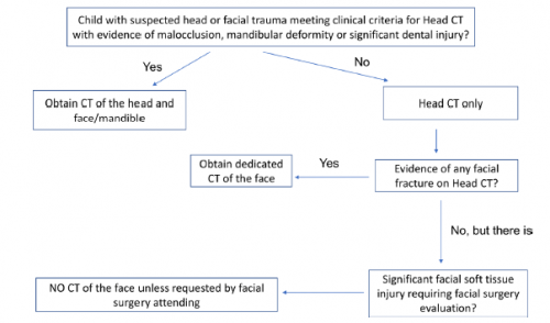
Here is the algorithm the authors recommend. Although designed for children, it should work for adults just as well.

Reference: Clinical and radiographic predictors of the need for facial CT in pediatric blunt trauma: a multi-institutional study. Trauma Surg Acute Care Open 2022;7:e000899.

How To Detect Bucket-handle Intestinal Injuries With CT Scan

A bucket-handle injury is a relatively uncommon complication of blunt trauma to the abdomen. It only occurs in a few percent of patients, but is much more likely if they have a seat belt sign.  The basic pathology is that the bowel mesentery (small bowel of sigmoid colon) gets pulled away from the intestinal wall.

This injury is problematic because it may take a few days for the bowel itself to die and perforate. Patients with no other injuries could potentially be discharged from the hospital before they become overtly symptomatic, leading to delayed treatment.

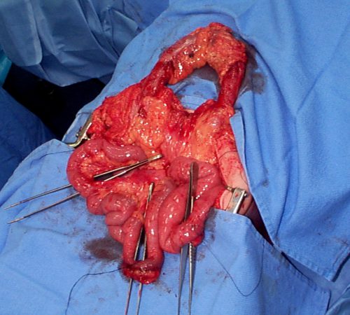
Here’s an image from my personal collection with not one, but four bucket-handle injuries.

Typical patients with suspected blunt intestinal injury are observed with good serial exams and a daily WBC count. If this begins to rise after 24 hours, there is a reasonable chance that this injury is present.

CT scan has not really been that reliable in past studies. There may be some “dirty mesentery”, which is contused and has a hematoma within it. But without a more convincing exam, it is difficult to convince yourself to operate immediately on these patients.

A paper was published by a group of radiologists at Duke University. It appears to be a case report disguised as a descriptive paper. It looks like they picked a few known bucket-handle injuries from their institution and back-correlated them with CT findings.

The authors called out the usual culprits:

  • Fluid between loops of bowel
  • Active bleeding in the mesentery
  • Bowel wall perfusion defects

But they also noted that traumatic abdominal wall hernias were highly with the injury as well. These are rare, but should bring intestinal injury to mind when seen.

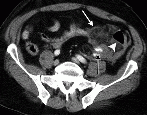
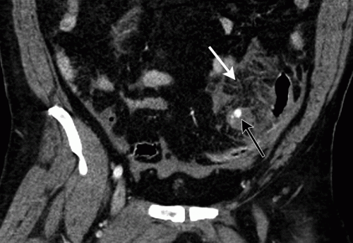
With newer scanners, radiologists are better able to detect subtle areas of hypoperfusion as well. This is a fairly good indicator of injury, especially when adjacent bowel appears normally perfused. Here are two examples. The black arrows denote active extravasation, and the white ones an area of hypoperfusion.

The authors add bowel wall hypoperfusion as another finding that may point to a bucket-handle type injury

Bottom line: Hold the phone! Don’t change your practice yet. This paper is not able to demonstrate how good this radiographic sign is. Looking at other radiology literature, the specificity is about 90%. But remember, that means that if they don’t have the CT finding, that’s true only 90% of the time.

Unfortunately the sensitivity is only 10%. So if you see it on the scan, they’ve got a 1 in 10 chance of actually having the injury. That’s not good enough for me to run to the operating room.

Here’s what I recommend: if your patient has an unconcerning exam and any of the usual culprits (pelvic fluid, inter-loop fluid, dirty mesentery, thickened bowel loops, abdominal wall hernia), perform serial exams and get a WBC the next morning. If the exam worsens, operate. If the WBC rises, consider laparoscopy to see if you need to make a bigger incision. And if you see this new kid on the block, the hypoperfused bowel, consider laparoscopy right away. 

I’m sure the radiologists and the technology will keep getting better. But for now, blunt intestinal injury still requires patience, perceptiveness, and a little luck.

References:

  • CT findings of traumatic bucket-handle mesenteric injuries. Am J Radiol 209:W360-@364, 2017.
  • Multidetector CT of blunt abdominal trauma. Radiology 265(3):678–693, 2012.

What? Still Using MRI For Cervical Spine Clearance?

Cervical spine clearance as evolved considerably over the years. First, there were five views of the spine using plain radiography. Then there were three. Then we moved to CT scan with clinical clearance. And currently, many institutions are relying only on CT.

But MRI has been used as an adjunct for quite some time. Initially, it was the tie breaker in patients who had equivocal CT findings, and for a while it was used for clearance in obtunded patients. And thanks to conflicting literature and disparate studies, the occasional usage became more frequent.

The group at Cedars-Sinai Medical Center in Los Angeles  noted that the percentage of patients undergoing MRI for cervical spine evaluation at their center slowly slowly crept up from 0.9% to 5.6% over a 10 year period. They designed a study to analyze the utility of this practice and inform their future practice.

Here are the factoids:

  • Over 9,000 patients had cervical spine CT during the 10-year study period; 513 (5.6%) were positive
  • Of the 513 CT-positive patients, 290 (56%) underwent an MRI. This showed:
    • Confirmation of the major injury in 250
    • Minor injury in 40
    • Clinically significant injury was seen in only 2 which was no surprise since they both had neurologic deficits
  • Of the 8,588 CT-negative patients, only 9 had clinically significant findings and 8 of them had neurologic deficits

Bottom line: So what have we learned here? First, MRI usage at Cedars-Sinai increased over time but was really not that useful. The main use was for imaging obtunded patients or those with an obvious neurologic deficit.

More than half of patients with positive CT scans also underwent MRI. If a major injury was seen on CT, MRI confirmed it. But if the CT findings were minor, none of the MRIs added any clinically significant findings in the absence of a neurologic deficit.

And what about MRI after negative CT? In the absence of a deficit, only one had a clinically significant finding (which only required a brace).

This study shows the wisdom of monitoring “how we do it.” There is sometimes some creepage away from what the literature shows is the best practice. The best way to remedy this is to do a good study, just like the authors did. They saw a slow change in practice, investigated it, and found that there was no good clinical reason for it. This gives the trauma program the ammunition to squelch the unwelcome behavior and return the clinicians to best practices.

Reference: Is MRI becoming the new CT for cervical spine clearance? Trends in MRI utilization at a Level I trauma center. J Tra publish ahead of print, DOI: 10.1097/TA.0000000000002752, 2020.

CT Contrast Via Intraosseous Catheter

The standard of care in vascular access in trauma patients is the intravenous route. Unfortunately, not all patients have veins that can be quickly accessed by prehospital providers. Introduction of the intraosseous device (IO) has made vascular access in the field much more achievable. And it appears that most fluids and medications can be administered via this route. But what about iodinated contrast agents via IO for CT scanning?

Physicians at Henry Ford Hospital in Detroit published a case report on the use of this route for contrast administration. They treated a pedestrian struck by a car with a lack of IV access sites by IO insertion in the proximal humerus, which took about 30 seconds. They then intubated using rapid sequence induction, with drugs injected through the IO device. They performed full CT scanning using contrast injected through the site using a power injector. Images were excellent, and ultimately the patient received an internal jugular catheter using ultrasound. The IO line was then discontinued.

This paper suggests that the IO line can be used as access for injection of CT contrast if no IV sites are available. Although it is a single human case, a fair amount of studies have been done on animals (goats?). The animal studies show that power injection works adequately with excellent flow rates.

The authors prefer using an IO placement site in the proximal humerus. This does seem to cause a bit more pain, and takes a little practice. A small xylocaine flush can be administered to reduce injection discomfort in awake patients. Additionally, the arm cannot be raised over the head for the torso portion of the scan.

Bottom line: CT contrast can be injected into an intraosseous line (IO) with excellent imaging results. Insert the IO in a site that you are comfortable with. I do not recommend power injection at this time. Although the marrow cavity can support it, the connecting tubing may not. Have your radiologist hand-inject and time the scan accordingly. And don’t be surprised if your radiology department doesn’t have a protocol for this!

Note: long term effects of iodinated contrast in the bone marrow are not known. For this reason, and because of smaller marrow cavities, this technique is not suitable for pediatric patients.

Reference: Intraosseous injection of iodinated computed tomography contrast agent in an adult blunt trauma patient. Annals Emerg Med 57(4):382-386, 2011.