Tag Archives: complications

Air Embolism From an Intraosseous (IO) Line

Intraosseous (IO) lines are a godsend when we are faced with a patient who desperately needs access but has no veins. The tibia is generally easy to locate and the landmarks for insertion are straightforward. They are so easy to insert and use, we sometimes “set it and forget it”, in the words of infomercial guru Ron Popeil.

But complications are possible. The most common is an insertion “miss”, where the fluid then infuses into the knee joint or soft tissues of the leg. Problems can also arise when the tibia is fractured, leading to leakage into the soft tissues. Infection is extremely rare.

This photo shows the inferior vena cava of a patient with bilateral IO line insertions (black bubble at the top of the round IVC).

During transport, one line was inadvertently disconnected and probably entrained some air. There was no adverse clinical effect, but if the problem is not recognized and the line is not closed properly, there could be.

Bottom line: Treat an IO line as carefully as you would a regular IV. You can give anything through it that can be given via a regular IV: crystalloid, blood, drugs. And even air, so be careful!

Top 10 Worst Complications: #1 Nasocerebral Tube

Minor complications from nasogastric tube insertion occur relatively frequently. Emesis is fairly common when the gag reflex is stimulated by the tube in the back of the oropharynx. An infrequent but possibly fatal one is insertion through the cribriform plate. 

The cribriform plate is located directly posterior to the nares and is part of the ethmoid bone. It is very porous in nature and weaker than the surrounding portions of the ethmoid. It is easily fractured, and can be seen is association with basilar skull fractures. This is one source for rhinorrhea in patients with these fractures.

Cribriform fracture is a contraindication to unprotected insertion of a nasogastric tube. If you look at the sagittal section below, the plate lies directly behind the nares. When inserting the NG tube, we are usually taught to aim the tube straight back. Unfortunately, this aims it directly at the cribriform. If a fracture is present, it is possible that you may be inserting a nasocerebral tube!

Cribriform plate - sagittal section

The usual symptoms when this occurs consist of immediate neurologic deterioration to coma, and a unilateral or bilateral blown pupil. The tube must not be withdrawn, because it will cause significant injury to the base of the brain. A stat neurosurgical consultation must be obtained, and if the patient is salvageable, the tube must be withdrawn through a craniectomy.

To avoid this dreaded complication, identify patients at risk for cribriform injury. They are:

  • patients with signs of trauma from eyebrows to zygoma
  • comatose patients
  • patients with signs of basilar skull fracture (Battle’s sign, raccoon eyes, oto- or rhinorrhea)

If your patient is at risk, follow these guidelines:

  • first, does the patient really need a gastric tube?
  • if comatose, insert an orogastric tube
  • if awake, don’t put the tube in their mouth, as they will gag continuously. Instead, place a lubricated, curved nasal airway. Then lube up a slightly smaller Salem sump tube and pass it through the airway.

Complications After Single-Look Laparotomy

Damage control laparotomy (DCL) has been around now for over 25 years. Many, many papers have been written on its benefits, and the decreased mortality for abdominal trauma specifically. In fact, its use has been generalized to trauma for all other body cavities as well.

However, with this improved mortality came an increase in complications. Incisional hernias remain common, as do episodes of delayed small bowel obstruction. Much of the emphasis in traumatic damage control surgery has now shifted to finding ways to close wounds more quickly and reduce the overall complication rate.

In contrast to damage control laparotomy, much less is known about the potential complications associated with the single-look trauma laparotomy.

This procedure is carried out more frequently than DCL, but we have spent less time studying outcomes and risk factors for complications in this group of patients.

The surgery group at Scripps Mercy Hospital in San Diego conducted a statewide retrospective review of a hospital discharge database of adult trauma patients over an eight-year period. Patients with multiple laparotomies were excluded, as it was assumed that these were damage control patients.

The primary outcomes studied were surgical complications, including bowel obstruction, hernia, fistula, wound infection or dehiscence, and evisceration. Complications were recorded during the initial admission, and during any readmissions in the study period.

Here are the factoids:

  • Over 3700 patients were identified as undergoing trauma laparotomy during the study period
  • About 2100 were left for review after excluding those with multiple laparotomies (DCL) or an unclear trauma mechanism
  • 80% of patients were male and 60% had a penetrating mechanism
  • One third of patients were readmitted for a surgery-related complication: SBO 18%, hernia 12%, infection 9%
  • Median time to readmission was about 4 months (range 1 week to 1.5 years)
  • Patients with blunt injury tended to present with complications earlier (6 days) than penetrating injuries (6 weeks)

Bottom line: This paper is unique in that it is one of the few that was able to follow a large patient population for complications occurring both during and after the initial admission. The overall complication rate was surprisingly high (33%), which is similar to that seen after emergency surgery.

Knowing all of this, what should we do? To date, we have not come close to solving the problems of postop adhesive small bowel obstruction, wound infection, and incisional hernia in any surgical population. However, this work points out the importance of counseling our patients about the potential for complications, how to recognize them, and when to present for evaluation and treatment.

Reference: Outcomes after single-look trauma laparotomy: A large population-based study. J Trauma 86(4):565-572, 2019.

Are Transfusing Too Much Blood During The MTP?

The activation of the massive transfusion protocol (MTP) for hypotension is commonplace. The MTP provides rapid access to large volumes of blood products with a simple order. Trauma centers each design their own protocol, which usually includes four to six units of PRBC per MTP “pack.”

This rapid delivery system, coupled with rapid infusion systems, allows the delivery of large volumes of blood and other blood products very quickly. But could it be that this system is too slick, and we are a bit too zealous, and could even possibly transfuse too much blood?

The trauma group at Cedars-Sinai in Los Angeles retrospectively reviewed their own experience via registry data with their MTP over a 2.5 year period for evidence of overtransfusion. All patients who received blood via the MTP were included. Patients who had a continuous MTP > 24 hours long, those who died within 24 hours, and those who had a missing post-resuscitation hemoglobin (Hgb) were excluded.

The authors arbitrarily defined overtransfusion as a Hgb > 11 at 24 hours. They also compared the Hgb at the end of the MTP and upon discharge with this threshold. They chose this Hgb value because it allows for some clinical uncertainty in interpreting the various endpoints to resuscitation.

Here are the factoids:

  • 240 patients underwent MTP during the study period, but 100 were excluded using the criteria above, leaving 140 study patients
  • Average injury severity was high (24) and 38% suffered penetrating injury
  • Median admission Hgb was 12.6
  • At the conclusion of the MTP, 71% were overtransfused using the study definition, 44% met criteria 24 hours after admission, and 30% did at time of discharge
  • Overtransfused patients were more likely to have a penetrating mechanism, lower initial base excess, and lower ISS (median 19)

The authors concluded that overtransfusion is more common than we think. This may lead to overutilization of blood products, which has become much more problematic during the COVID epidemic. They recommend that trauma centers track this metric and consider it as a quality of care measurement.

Bottom line: This is a nicely crafted and well-written study. It asks a simple question and answers it with a clear design and analysis. The authors critique their own work, offering a comprehensive list of limitations and a solid rationale for their assumptions and conclusions. They also offer a good explanation for their choice of Hgb threshold in defining overtransfusion.

I agree that overtranfusion truly does occur, and I have seen it many times first-hand. The most common reason is the lack of well-defined and reliable resuscitation endpoints. How do we know when to stop? What should we use? Blood pressure? Base excess? TEG or ROTEM values? There are many other possibilities, but none seem reliable enough to use in every patient. 

Patients with penetrating injury proceeding quickly to OR more commonly experience overtransfusion. This may be due to the reflexive administration of everything in each cooler and the sheer speed with which our rapid infuser technology can deliver products. The more product in the cooler, the more that is given, which may lead to the overtranfused condition. 

The authors suggest reviewing the makeup of the individual MTP packs, and this makes sense. Are there too many in it? This could be a contributing factor to overtransfusion. It might be an interesting exercise to do a quick registry review at your own center to obtain a count of the number of MTP patients with a final Hgb > 11. If you find that your numbers are high, consider reducing the number of red cell packs in the cooler to just four. But if you already only include four, don’t reduce it any further. And in any case, critically review the clinical indicators your  surgeons use to decide to end the MTP to see if, as a group, they can settle on one to use consistently. 

Reference: Overtransfusion of packed red blood cells during massive transfusion activation: a potential quality metric for trauma resuscitation. Trauma Surg Acute Care Open 7:e000896., July 26 2022.

Surveillance For Splenic Pseudoaneurysm After Injury

When it comes to repeat CT scanning after splenic injury, there are believers and there are non-believers. In my experience, the majority of centers in the US are non-believers. However, there is a new paper in press that attempts to convince us that more should become believers.

I think the biggest lesson to be learned from this paper is that WE SHOULD READ THE ENTIRE PAPER before drawing conclusions. I have said this in the past and I will say it again. In this case, not only did I read the entire paper, but I had to dig deep into the references it cited as well.

Nonoperative management of splenic injuries has a very high success rate if done properly. Some papers claim this can be up to 93%, which parallels my experience. This success rate involves excluding unstable patients (they need to be in the operating room) and planned use of angioembolization in select patients. Over the years we have found that we need to do less and less in the management of solid organ injury patients:

  • No bedrest
  • No starvation (NPO status)
  • No serial blood draws
  • No repeat CT scan
  • Few limitations on activity after discharge

For an example of a practice guideline that demonstrates that less is more, use the download link at the end of this post.

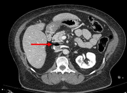
But back to the question about repeat CT scanning before discharge. Why do we need to do this? The usual reason is that “we want to find delayed pseudoaneurysms.” And why is that important? “It might bleed!”

Really? Let’s look into that through the lens of this new paper by the group at the University of Cincinnati. They performed a retrospective study of their experience with patients who had sustained blunt splenic injury during a recent three-year period. They were interested in how many underwent splenectomy or splenorrhaphy, who had repeat CT imaging, who went to interventional radiology (IR) and when, and which ones were found to have pseudoaneurysms and what was done about it.

Here are the factoids:

  • There were 539 patients who met inclusion criteria, with an average ISS of 24
  • Of these, 46 died during their hospital stay (none from their splenic injury)
  • Focusing on the 248 patients with higher grade injuries (III-V), 125 (50%) underwent emergent or delayed splenectomy. Early vs late operation was not broken out, but this is a startlingly high number!
  • Of the higher grade injured patients who kept their spleens, 97% underwent repeat CT around day 5
  • Delayed pseudoaneurysms were detected in the following patients:
    • Grade III: 10 of 88 patients (11%). Then 8 of those 10 went to IR, and 5  of 10 had splenectomy!
    • Grade IV: 7 of 24 (29%).  Then 8 of the 7 (error in the paper?) went to IR and 3 of 7 had splenectomy!
    • Grade V: 2 of 5 (40%). Both of these patients went to IR and somehow kept their spleens.

The authors conclude that routine followup CT imaging identifies splenic pseudoaneurysms allowing for interventions to minimize delayed complications.

Bottom line: Whoa! There’s a lot going on here. My first observation is that this center does a lot of splenectomies! Of the 539 patients (all comers) who were included in the study, 129 (24%)  lost their spleens. If grade I-II injuries are excluded that percent rises to 50%!

Only eight splenectomies were performed after the repeat CT. This would imply that there were either a lot of unstable patients with splenic injury, the institutional indications for this procedure arbitrarily include grade, or there is a lot of variability in the decision to perform it.

I think there are really two questions to answer here. 

  1. Does delayed splenic pseudoaneurysm occur? The answer is yes. There are a few studies (performed by believers) that demonstrate new pseudoaneurysms after repeat CT. I’m convinced.
  2. Do we care? The real question is, do these pseudoaneurysms cause harm? The fear is that they might explode at some point after patient discharge and cause a major problem.

Papers written by the believers cite a number of old studies and give numbers between 2% and 27% for incidence of delayed hemorrhage. Well, I tracked down all of these papers, including the ones they cited. And it doesn’t add up.

  • One paper from a believer institution found no delayed bleeds.
  • Several papers were for pediatric patients, whose spleens don’t behave like adult ones. They found one case after discharge in one out of 276 patients across three studies.
  • Of 76 adolescents, none encountered delayed bleeds

Many of the papers cited regarding bleeding complications are very old. CT scanners had less resolution, and in many papers, IR was not even a consideration. 

So here’s my take. Yes, delayed pseudoaneurysms occur. In children we don’t care. They almost never cause a problem. But in adults, they can and do cause issues and should be embolized shortly after the initial scan. 

Once embolized, the ones seen on that initial scan are effectively neutralized and do not need a repeat scan. The small ones that might pop up later may very well be part of the healing process. And they may not even occur if angioembolization is done early. It seems unlikely that anything further is needed.

But remember, clinical judgement trumps all. If your patient starts complaining of new abdominal symptoms while in the hospital or after discharge, get a prompt CT scan to rule out any developing complications.

Sample solid organ injury protocol: click here

Reference: Delayed splenic pseudoaneurysm identification with surveillance imaging. J Trauma Acute Care Surg. 2022 Mar 22. doi: 10.1097/TA.0000000000003615. Epub ahead of print. PMID: 35319540.