Tag Archives: blush

Natural History of the Splenic Blush

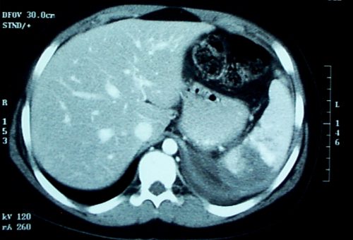
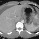
In my last post, I described the two types of solid organ “blushes.” I also described my thoughts on the natural history of these findings. Now, a multicenter study on the natural progression of the splenic “blush” has just been published. I found this paper very interesting, because it challenged some of my own existing beliefs. But once I read it, my enthusiasm faded.

The Western Trauma Association sponsored a multicenter (17 Level I and II centers) review of data collected prospectively over an unspecified period of time. Patients were excluded if their injury was older than 24 hours, if they had a previous splenic injury, and if they had any number of diseases or hereditary conditions that might affect the spleen. Strict definitions of nonbleeding and actively bleeding injuries were applied, and detailed information on intervention and outcomes was collected.

Here are the factoids:

  • 200 patients were enrolled from 17 centers, but the paper does not state how long that took
  • 20% were low grade (1 or 2) and 80 % high grade (3-5)
  • 29% had a pseudoaneurysm, and 83% showed extravasation, which means that several patients had both
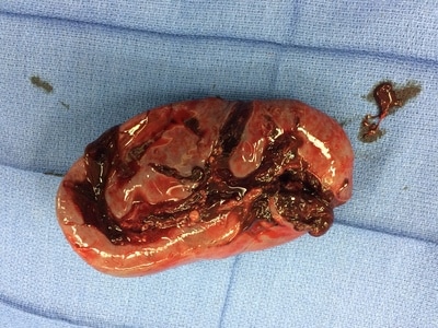
  • 15% underwent early splenectomy, 59% underwent angiography, and 26% were observed
  • For those with initial angiography, 6% had repeat angio and 7% eventually underwent splenectomy
  • Of those were were initially observed, 9% had delayed angio and 8% underwent splenectomy
  • Based on a read by an expert radiologist, an actively bleeding injury was associated with a 41% splenectomy rate
  • The authors conclude that the majority of patients with spleen injury with pseudoaneurysm or extravasation are managed with angio and embolization and that splenectomy remains a rare event (??)

Bottom line: This paper just doesn’t do it for me. The biggest problem is that it is what I call a “we do it the way we do it” study. It examines how 17 different centers evaluate and treat patients with significant splenic injury. There was no guidance or guideline on how to treat, so they each did it their way. And the number of patients was small.

They don’t tell us anything about the use or effectiveness of angio by grade. Or whether the specific hospitals routinely rely on angio rather than just going to the OR for high grade injuries (typically if angio response times are long).

Unfortunately, this paper gives the appearance of containing a lot of interesting stuff. But a 15% initial splenectomy rate is not a “rare event” in my book. Everything published here is at odds with what I’ve observed over the years for centers with well developed management guidelines and easy access to angio (< 5% splenectomy rate in hemodynamically stable patients with nonoperative management).

My recommendation is to send all stable patients with pseudoaneursym and/or extravasation to angio immediately! Yes, some will have nothing found by the time they get to angio, and you’ll have to come up with a plan at that point. But most have something wrong, and it won’t stop until it’s been plugged up (or your patient bleeds to death, whichever comes first)!

This article has all the right buzzwords: multicenter, prospective data, etc. But it’s already been moved to my recycle bin. 

Related post:

Reference: Natural history of splenic vascular abnormalities after blunt injury: A Western Trauma Association multicenter trial. J Trauma 83(6):999-1005, 2017.

Splenic Vascular Blush

Contrast blush is always a concern when seen on CT of the abdomen for trauma. It can represent one of two things, and both are bad:

  • Active extravasation of contrast
  • Splenic pseudoaneurysm

These two clinical issues can be distinguished by looking at the location of the contrast and its persistence. A pseudoaneurysm is located within the parenchyma, and the contrast will wash away, so it will not be visible on delayed images. Contrast that extends beyond the parenchyma or persists in delayed views represents active bleeding. In either case, the failure rate of nonoperative management exceeds 80% in adults without additional measures being taken.

Clinically, these patients usually act as if they are losing volume and require additional crystalloid and/or blood transfusion. The natural history in adults is for bleeding to continue or for the pseudoaneurysm to rupture, resulting in a quick trip to the operating room.

If vital signs can be maintained with fluids and blood, a trip to interventional radiology may solve the problem. Selective or nonselective embolization can be carried out and patients with only a few bleeding points can be spared operation. However, if multiple bleeding areas are seen, it is probably better to head to the OR for splenorrhaphy or splenectomy.

The image below shows likely areas of extravasation. They are a bit large to be pseudoaneurysms.

Spleen Blush-CT

Children are different than adults. Extravasation from spleen injuries in prepubescent children frequently stops on its own. Angiography should only be used if the child is failing nonoperative management.

Next post: A new paper looks at the natural history of these lesions.

Contrast Blush in Children

A contrast blush is occasionally seen on abdominal CT in patients with solid organ injury. This represents active arterial extravasation from the injured organ. In most institutions, this is grounds for call interventional radiology to evaluate and possibly embolize the problem. The image below shows a typical blush.

Splenic contrast blush

This thinking is fairly routine and supported by the literature in adults. However, it cannot be generalized to children!

Children have more elastic tissue in their spleen and tend to do better with nonoperative management than adults. The same holds true for contrast blushes. The vast majority of children will stop bleeding on their own, despite the appearance of a large blush. In fact, if children are taken to angiography, it is commonplace for no extravasation to be seen!

Angiography introduces the risk of local complications in the femoral artery as well as more proximal ones. That, coupled with the fact that embolization is rarely needed, should keep any prudent trauma surgeon from ordering the test. A recently released paper confirms these findings.

The only difficult questions is “when is a child no longer a child?” Is there an age cutoff at which the spleen starts acting like an adult and keeps on bleeding? Unfortunately, we don’t know. I recommend that you use the “eyeball test”, and reserve angiography for kids with contrast extravasation who look like adults (size and body habitus).

Reference: What is the significance of contrast “blush” in pediatric blunt splenic trauma? Davies et al. J Pediatric Surg 2010 May; 45(5):916-20.

Delayed Splenic Rupture: Part 2

Yesterday I wrote about the history of “delayed splenic rupture.” Today I’ll discuss how to deal with it.

If possible, try to avoid ever having to mess around with this clinical problem. If you order an abdominal CT after blunt trauma and see a splenic contrast blush of either type (pseudoaneurysm or extravasation, see left photo), then deal with it before the patient even knows he has a problem. A trip to interventional radiology will usually solve the problem. And if embolized, these patients almost never come back with a bleeding problem.

As I’ve said many times before, if the patient is hemodynamically compromised, then an OR visit is required. The usual solution is splenectomy. Some recommend repairing the spleen, but this is technically more difficult than it sounds, and it is difficult for the surgeon to sleep soundly after performing one of these.

Lets say you inherited one of these from someone else, or ignored the warning signs on the initial CT. The usual time frame for presentation to the ED with acute bleeding is 7 to 10 days after the initial injury. If they are not stable, physical exam or FAST will quickly direct you to the OR, once again for splenectomy. Some patients will stabilize with fluids and can safely be sent to CT scan.

Once the CT confirms what the problem is, a trip to interventional radiology is in order if the patient remains stable. Here is the key: the radiologist must embolize something! If they find a bleeding vessel, then they can selectively embolize it. If they don’t, then the main splenic artery should be embolized. This will decrease the arterial pressure head, but won’t eliminate it. It will decrease the likelihood of additional bleeding as much as possible.

At this point, the patient should be admitted to the trauma service and monitored using your solid organ injury protocol. If they have any hemodynamic issues, it’s time to remove the spleen. Remember, this is the third time they’ve had a problem, and like in baseball, their spleen is out! Attempted splenorrhaphy at this point is pointless and may lead to yet another operation.

Related posts:

Delayed Splenic Rupture: Part 1

This post was prompted by a paper that somehow got into the Journal of Trauma this month on nonoperative management of delayed splenic rupture after trauma. It’s a bad retrospective review of 15 patients which I’ll say more about tomorrow. There’s very little good literature on this topic, so I wanted to share some personal observations.

Back in the days before CT scan (and unfortunately, I remember them), the diagnosis of abdominal injury was much more difficult. It was primarily qualitative, meaning that we somehow figured out that they either had it or they didn’t. We could not very easily figure out what specific injuries a given patient had. However, management was simple: we went to the operating room, found out and fixed it.

Sometimes, though, we would encounter a patient who had been involved in some type of blunt trauma a week or two earlier who presented to the ED with left-sided abdominal pain, shock and anemia. The diagnosis was “delayed splenic rupture” and they were taken to OR for a splenectomy.

When CT scan came along, we found out that these were actually “delayed recognition of splenic injury.” We still took them to the OR for splenectomy, but with experience this slowly gave way to splenic repair, and then to nonoperative management. 

There is still one subset of these injuries that is problematic: spleen injury with a contrast blush. It turns out that there are really two types of blush: contrast seen within a pseudoaneurysm within the splenic pulp, and extravasation. And furthermore, the pseudoaneurysm is the culprit in most “delayed splenic ruptures.”

Tomorrow, I’ll write about how to recognize this potential problem, what to do about it acutely, and what to do if it was missed and the patient presents to your ED ten days later in shock.

Related post:

Reference: Nonsurgical management of delayed splenic rupture after blunt trauma. J Trauma 72(4):1019-1023, 2012.