Tag Archives: blunt trauma

Best Of EAST 2023 #5: Imaging The Elderly

Several papers have been published over the years regarding underdiagnosis when applying the usual imaging guidelines to elderly trauma patients. Unfortunately, our elders are more fragile than the younger patients those guidelines were based on, leading to injury from lesser mechanisms. They also do not experience pain the same way and may sustain serious injuries that produce no discomfort on physical exam. Yet many trauma professionals continue to apply standard imaging guidelines that may not apply to older patients.

EAST sponsored a multicenter trial on the use of CT scans to minimize missed injuries. Eighteen Level I and Level II trauma centers prospectively enrolled elderly (age 65+) trauma patients in the study over one year. Besides the usual demographic information, data on physical exams, imaging studies, and injuries identified were also collected. The study sought to determine the incidence of delayed injury diagnosis, defined as any identified injury that was not initially imaged with a CT scan.

Here are the factoids:

  • Over 5,000 patients were enrolled, with a median age of 79
  • Falls were common, with 65% of patients presenting after one
  • Nearly 80% of patients actually sustained an injury (!)
  • Head and cervical spine were imaged in about 90% of patients, making them the most common initial studies
  • The most commonly missed injuries involved BCVI (blunt carotid and vertebral injury) or thoracic/lumbar spine fractures
  • 38% of BCVI injuries and 60% of T/L spine fractures were not identified during initial imaging
  • Patients who were transferred in, did not speak English, or suffered from dementia were significantly more likely to experience delayed diagnosis

The authors concluded that about one in ten elderly blunt trauma patients sustained injuries in body regions not imaged initially. They recommended the use of imaging guidelines to minimize this risk.

Bottom line: Finally! It has taken this long to perform a study that promotes standardizing how we perform initial patient imaging after blunt trauma. Granted, this study only applies to older patients, but the concept can also be used for younger ones. The elderly version must mandate certain studies, such as head and the entire spine. Physical exams can  still be incorporated in the guidelines for younger patients but not the elderly.

The overall incidence of BCVI was low, only 0.7%. But its presence was missed in 38% of patients, setting them up for a potential  stroke. Some way to incorporate CT angiography of the neck will need to be developed. The risk / benefit ratio of the contrast load vs. stroke risk will also have to be determined.

Here are my questions and comments for the presenter/authors:

  • Did you capture all of the geriatric patients presenting to the study hospitals? By my calculation, 5468 patients divided by 18 trauma centers divided by 14 months of study equals 22 patients enrolled per center per month. Hmm, my center sees more than that number of elderly injured patients in the ED per day! Why are there so few patients in your study? Were there some selection criteria not mentioned in the abstract?
  • Why should we believe these study numbers if you only included a subset of the total patients that were imaged?

My own reading of the literature leads me to believe that your conclusions are correct. I believe that all centers should develop or revise their elderly imaging guidelines to include certain mandatory scans regardless of how benign the physical exam appears. Our elders don’t manifest symptoms as reliably as the young. But the audience needs a little more information to help them understand some of the study numbers.

Reference: SCANNING THE AGED TO MINIMIZE MISSED INJURY, AN EAST MULTICENTER TRIAL. EAST 2023 podium abstract #12.

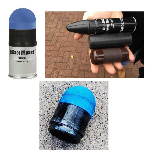
What Is: A Rubber Bullet?

The protests in cities across the country continue. Many are peaceful, but not all. In some cases, police have resorted to “non-lethal” weapons to control and disperse crowds.

Although these weapons are called non-lethal, that’s not entirely true. The projectiles, gases, and powders that are being used all have some degree of morbidity and mortality. They are certainly less so than traditional projectiles (bullets), but serious and fatal injures can and do occur.

One item that is talked about in the news is the rubber bullet. What are these, exactly? The generic term is a “kinetic impact projectile” (KIP). It encompasses a variety of objects that are not designed to penetrate flesh like a regular bullet. They can be bullets, beanbags, sponges, pellets, and other odds and ends.

And the so-called “rubber bullet” isn’t even necessarily made of rubber. It can be plastic, metal, rubber, or other substances.

There is very little published data on injuries caused by KIPs. Because of their odd shapes, they tend to tumble when they are fired. This decreases aiming accuracy substantially when the target is distant. They are designed to be aimed at the lower extremities. However, if the aim is too high or the round is fired at close range, it can be lethal.

Here are some typical injuries that have been descirbed:

  • Subdural and intraparenchymal hematomas
  • Skull and facial fractures
  • Eye injuries leading to blindness (this happened to a photographer in Minneapolis last week)
  • Rib fractures and pulmonary contusions
  • Spleen laceration
  • Blunt intestinal injury

Here’s a video from one of the manufacturers that shows the amount of target deformation caused by a sponge tipped bullet. Very impressive.

YouTube player

(Tumblr viewers please click here to view video)

Bottom line: Although they sound relatively innocuous, kinetic impact projectiles of any kind are far from it. If you are called to treat a patient who has been shot with one, be sure to do a very thorough evaluation. Most head, neck, and torso injuries should undergo CT scanning to delineate deep or occult injuries in detail. In-hospital observation of torso injuries is warranted, as well as a good tertiary exam.

AAST 2019 #2: Predicting Abdominal Operation After Blunt Trauma – The RAPTOR Score

Patients with blunt abdominal injury, particularly those with seat belt signs, can be diagnostically very challenging. If the patient is stable and does not have peritonitis, CT scan is typically the first stop after the trauma resuscitation room. As many trauma professionals know, the radiographic findings can be subtle and/or not very convincing.

The trauma group at the University of Tennessee in Memphis sought to identify specific findings that might help us better identify patients that will need laparotomy. They retrospectively identified all their mesenteric injuries over a five-year period. A single blinded radiologist (is this an oxymoron or not?) reviewed all 151 patient images who underwent laparotomy, looking for predictors of bowel or mesenteric injury.  All of the predictors were then converted into a scoring system called RAPTOR (radiographic predictors of therapeutic operative intervention; kind of a stretch?). These predictors were then subjected to multivariate regression analyses to try to tease out if there were any independent predictors of injury.

Here are the factoids:

  • A total of 151 patients were identified over the 5 year period; 114 underwent laparotomy
  • Of the 114 operated patients, two thirds underwent a therapeutic laparotomy and the other third were nontherapeutic
  • There no missed injuries in the non-operated patients
  • The components of the RAPTOR score were culled from all the potential findings, and were determined to be
    • Multifocal hematoma
    • Acute arterial extravasation
    • Bowel wall hematoma
    • Bowel devascularization
    • Fecalization (of what??)
    • Free air
    • Fat pad injury (??)
  • Linear regression then showed that only three of these, extravasation, bowel devascularization, and fat pad injury to be independent predictors of injury
  • If three or more RAPTOR variables were present, then the sensitivity, specificity, and positive predictive values for injury were 67%, 85%, and 86%, and an area under the receiver operating characteristic curve (AUROC) of 0.91

The authors concluded that the RAPTOR score provided a simplified approach to detect patients who might benefit from early laparotomy and not serial abdominal exams. They go further and say it could potentially be an invaluable tool when patients don’t have clear indications for operation.

It looks like there are two things going on here at the same time. First, a new potential scoring system is being piloted. And second, a regression analysis is being used to examine the data as well. 

But first, let’s back up to the beginning. This is a retrospective study, with a relatively small size. This makes it far harder to ensure that the results will be significant, or at least meaningful. Use of a single radiologist can also be problematic, especially since many of the CT findings with this mechanism of injury are subtle. 

The reported performance of the RAPTOR score is a bit weak. The listed statistics show that it accurately identified only two thirds of those who needed an operation and 85% of those who didn’t. The AUROC for the regression is very good, though. Could a good old-fashioned serial exam scenario be better?

Bottom line: It will be interesting to hear the background on RAPTOR vs regression, and find our how the authors will use or are using these tools.

Here are my questions for the presenter and authors:

  • Why did you decide to create a scoring system that uses a set of variables that may be dependent on each other? Isn’t the regression equation better?
  • Has this information changed your practice? It seems that the two of the three regression variables are fairly obvious reasons to operate (active extravasation and devascularization). Do you really need the rest?
  • Has this study helped you decrease the non-therapeutic laparotomy rate for blunt abdominal injury?
  • And please define fecalization and fat pad injury!

I’m looking forward to hearing this presentation!

Reference: RADIOGRAPHIC PREDICTORS OF THERAPEUTIC OPERATIVE INTERVENTION AFTER BLUNT ABDOMINAL TRAUMA: THE RAPTOR SCORE. AAST 2019 Oral Paper 6.


Practical Tip: Evaluation of Hematuria in Blunt Trauma

Bloody urine is a relatively uncommon finding in blunt trauma patients. Hematuria ranges from microscopic to gross. Microscopic means blood that can only be seen with a microscope, and gross means visible to the naked eye. In trauma, we only care about gross hematuria, which ranges from the faintest of pink to the deepest red.

In the picture above gross hematuria is present in all tubes but the far right one. Those four will need further evaluation.

In trauma, gross hematuria is a result of an injury to kidney, ureter or bladder. Blunt injury to the ureter is so rare it’s reportable, so you can pretty much forget that one unless the mechanism is extreme. So you really just need to focus on kidney and bladder.

Any victim of blunt trauma that presents with visible hematuria needs to be evaluated by CT of the abdomen and pelvis with an added CT cystogram. Standard CT technique is done without a urinary catheter, or with the catheter clamped. This is not acceptable for hematuria evaluation, as only 50% of bladder injuries show up with this technique.

CT cystogram is an add-on to the standard CT, and consists of the administration of contrast into the bladder which is then kept under pressure while the scan is performed. Delayed slices through the pelvis after the bladder is depressurized and emptied is routine. Nearly 100% of bladder injuries are detected using this technique.

If the CT shows a renal laceration or hematoma, the patient should be admitted and managed according to your solid organ injury protocol. Kidney injuries fare better that livers and spleens, and only rarely require surgery. If no kidney or bladder injury is seen, the default diagnosis of a renal contusion is the culprit. No treatment is needed, and the patient can be discharged if no other injuries are present. The blood will clear over a few days, but may disappear and reappear a few times in the process. Be sure to warn the patient that this may occur, or you may receive some surprise phone calls. The patient can followup with their primary care physician in a week or two.

The majority of these injuries do not require urologic consultation. Complex injuries with extravasation of urine out of the kidney, or injuries to the collecting system should be referred to a urologist, however.

EAST Guidelines: Blunt Traumatic Aortic Injury

The Eastern Association for the Surgery of Trauma (EAST) has been helping trauma professionals through the publication of practice guidelines for more than 15 years. Members of EAST donate their time to review reams of literature, good and bad, to try to determine the answers to common or puzzling clinical questions.

Why follow a practice guideline? Quite simply, if properly developed, a guideline represents our best understanding of the “correct” answer to the question posed. And as many of you who follow this blog already know, things that “seem to make sense” frequently are totally wrong. Your own experience is poignant, but the pooled experience of the many others who contributed to research on the topic in question is much more significant.

So on the the practice guideline for blunt traumatic aortic injury (BTAI). This one answers three questions. I will list each, followed by the conclusions reached through the literature review.

1. In patients with suspected BTAI, which diagnostic modality should be chosen: CT angiography of the chest, or conventional catheter angiography?

  – Catheter angiography was the standard for decades. When the first EAST guideline on this topic was released 15 years ago, CT angiography was only a level III recommendation because experience with it was lacking. CT technology has advanced rapidly, with multiple detectors, helical scanning, and incredible computing power. Although the quality of the evidence is somewhat low, the panel strongly recommends the use of CT angiography due to its ready availability, speed, low invasiveness, and ability to detect and define other injuries.

2. Should endovascular or open repair be selected in order to minimize stroke, renal failure, paraplegia, and death?

  – Once again, the quality of available data is so so. However, it was possible to detect differences in outcome in comparative studies. The panel strongly recommends the use of endovascular repair in patients who do not have contraindications due to its lower blood loss, mortality, and paraplegia, and equivalent risk of stroke. Furthermore, it is performed more frequently now than open repair, and experience is thus greater at many institutions, further reducing complications.

3. Should the repair be performed immediately or delayed in order to minimize stroke, renal failure, paraplegia, and death?

  – Literature review revealed that the incidence of renal failure and paraplegia were lower with delayed repair, while renal failure was the same in patients with significant associated injuries. There was benefit to delaying repair until resuscitation was achieved and any other life threatening injuries were addressed. The panel recommends that delayed repair be carried out once these other conditions have been corrected. The procedure should not be delayed until the next morning for the convenience of the surgeons.

Related posts:

Reference: Evaluation and management of blunt traumatic aortic injury: a practice guideline from the Eastern Association for the Surgery of Trauma. 78(1):136-146, 2015.